
Normal Brain MRI Report
Review a normal adult brain MRI example, sequence-dependent limits, and measured handling of age-related findings.
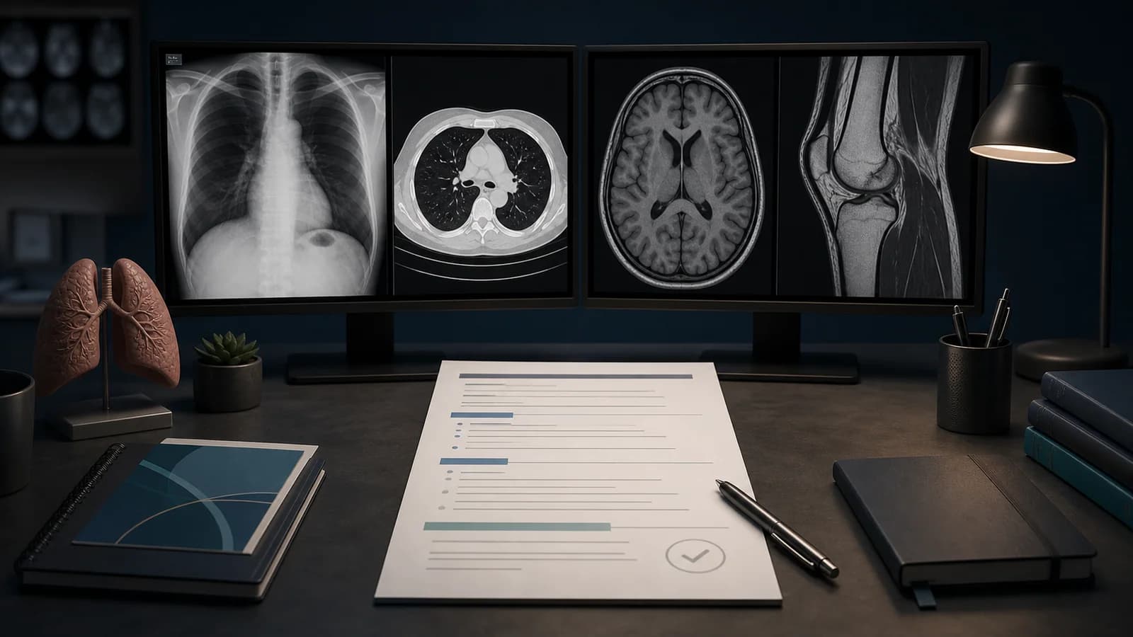
A normal brain MRI report should reflect the sequences used and the clinical question. A routine noncontrast study does not exclude small metastases, pituitary microadenoma, epileptogenic lesions, or cranial nerve disease as confidently as a targeted protocol. The normal template must preserve the actual scope of the examination.
This example is for routine adult brain MRI. White matter foci, volume loss, and vascular tortuosity can be age related and are not automatically pathologic. Interpret them with age, risk factors, symptoms, and prior imaging.
Assessment framework
- Survey cerebral and cerebellar parenchyma on T1, T2, and FLAIR images.
- Assess diffusion for acute ischemia and susceptibility images for blood products.
- Review ventricular size, sulci, and midline structures.
- Inspect brainstem, cerebellopontine angles, sella, and craniocervical junction.
- Check the orbits, paranasal sinuses, and mastoids within the acquired field.
What the report should contain
- State contrast use and core sequences in the technique section.
- Address parenchymal signal, diffusion, hemorrhage, mass, and mass effect separately.
- When ventricles are enlarged, interpret them with sulcal size.
- Do not label a few nonspecific FLAIR foci as definite demyelination.
- Do not close a targeted clinical question with a normal routine report.
Common errors
- Calling every white matter focus abnormal for age.
- Excluding enhancing disease on a noncontrast examination.
- Turning sinus or mastoid signal into an automatic diagnosis of infection.
Example report wording
Example impression: No intracranial mass, acute infarction, hemorrhage, or pathologic mass effect is identified. Ventricles and sulci are appropriate for age. Brain parenchymal signal is within normal limits. This conclusion applies to the routine noncontrast protocol; a pituitary, epilepsy, or cranial nerve question requires the appropriate targeted examination.
Verify the normal text against the images with the brain MRI reporting checklist.
References
- American College of Radiology. Practice Parameter for MRI of the Head and Neck. Source (opens in a new tab)
- American College of Radiology. Practice Parameter for CT of the Head. Source (opens in a new tab)
Clinical note: This material is for radiology education. Do not generate an automatic diagnosis or management decision without checking guideline scope, patient factors, and local policy.
Clinical use note
This content is educational and is not patient-specific medical advice. Every phrase, template, classification result, and recommendation must be verified and adapted by a physician using the complete examination, clinical context, current guidelines, and institutional protocol.
Adapt the report framework to the case
Use this review order with an editable RadPhrases template and verify every line against the images.
Related calculators
Browse the other checklists, phrase sets, and systematic approaches in this topic.