
Fleischner Society Thoracic Imaging Glossary 2024: Practical Terminology
A practical guide to applying the Fleischner Society's 2024 thoracic imaging terminology consistently in CT interpretation and radiology reports.
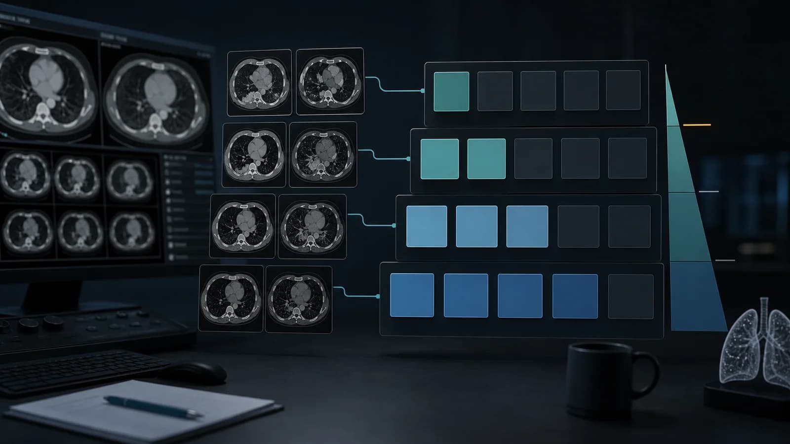
The 2024 Fleischner Society glossary standardizes the words used to describe thoracic anatomy, physiology, and imaging abnormalities. Its practical value is consistency: a descriptive term should communicate an observable imaging feature without silently implying an unproven diagnosis. Terms must still be interpreted in the clinical setting and in relation to acquisition technique.
Anatomic foundations
- The acinus is the gas-exchanging unit distal to a terminal bronchiole and is not normally resolved on CT.
- The secondary pulmonary lobule is the smallest unit outlined by connective-tissue septa; its center contains a bronchiole and pulmonary artery branch.
- Interlobular septa, fissures, pleural surfaces, and bronchovascular bundles provide the landmarks used to classify disease distribution.
- Small-airway disease is often inferred from indirect findings such as tree-in-bud nodules, mosaic attenuation, and expiratory air trapping.
Attenuation and air-space terms
- Ground-glass opacity is increased lung attenuation through which vessels and bronchial walls remain visible.
- Consolidation is increased attenuation that obscures underlying vessels and airway walls; an air bronchogram may remain visible.
- Mosaic attenuation describes patchwork regions of differing attenuation and may reflect small-airway, vascular, or infiltrative disease.
- Air trapping is failure of lung attenuation and volume to decrease normally on expiration; expiratory CT improves confidence.
Nodules and distribution
A pulmonary nodule is a focal, approximately rounded opacity measuring up to 30 mm; a larger lesion is termed a mass. State whether a nodule is solid, part-solid, or ground glass, and describe margins, calcification, cavitation, and growth. For diffuse nodules, identify a centrilobular, perilymphatic, random, or mixed distribution. Avoid using distributional language when the relationship to the secondary lobule cannot be established.
Airways, cysts, and fibrosis
- Bronchiectasis requires abnormal bronchial dilatation supported by features such as an increased bronchoarterial ratio, lack of tapering, or visible peripheral airways.
- Traction bronchiectasis or bronchiolectasis refers to airway dilatation caused by surrounding fibrosis and architectural distortion.
- A cyst is a circumscribed air- or fluid-containing space with a definable wall; bullae, blebs, pneumatoceles, and cavities should be named only when their defining context is present.
- Honeycombing consists of clustered, usually subpleural cystic air spaces in an established fibrotic pattern and should not be assigned from an isolated cystic focus.
Turning terminology into a report
- Begin with the dominant finding and its precise anatomic distribution.
- Use the glossary term for the observed feature, then separately state the favored diagnosis or differential.
- Document extent, severity, chronicity, and change from prior imaging.
- Use inspiratory, expiratory, prone, or contrast-enhanced acquisitions only when relevant to the question and note technical limitations.
- Avoid vague legacy terms such as “infiltrate” when a more specific descriptor is available.
- When findings overlap categories, describe the components rather than forcing a single label.
Clinical use note
This content is educational and is not patient-specific medical advice. Every phrase, template, classification result, and recommendation must be verified and adapted by a physician using the complete examination, clinical context, current guidelines, and institutional protocol.
Build the category and report statement together
Use the relevant RadPhrases calculator, then verify the result against the official guideline and the complete examination.
Related calculators
Browse the other checklists, phrase sets, and systematic approaches in this topic.