
Normal Contrast-Enhanced Abdomen and Pelvis CT Report
Use an editable normal contrast-enhanced abdomen and pelvis CT example while preserving phase, technical limits, and case-specific findings.
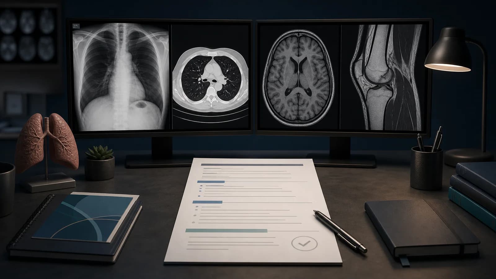
A normal CT template can serve as a checklist, but it cannot replace image review. Contrast phase, oral contrast, prior surgery, sex, and the clinical question change the text. Calling an unseen organ normal or excluding luminal disease beyond the technique reduces report reliability.
This structure is a teaching example for an adult diagnostic-quality contrast-enhanced CT of the abdomen and pelvis. Trauma, cancer staging, stone assessment, active bleeding, enterography, and vascular examinations require different protocols and impression language.
Assessment framework
- Assess liver, biliary system, pancreas, spleen, and adrenal glands.
- Review renal parenchyma, collecting systems, and bladder in the available contrast phase.
- Survey stomach, small bowel, colon, and appendix where visible.
- Inspect peritoneum, mesentery, retroperitoneum, and lymph nodes.
- Check aorta, major branches, portal veins, bones, and lung bases.
What the report should contain
- Technique: Contrast-enhanced CT of the abdomen and pelvis was obtained in the portal venous phase.
- Findings: No focal solid-organ lesion, biliary dilatation, or hydronephrosis is identified.
- Bowel: No obstruction or free air; luminal assessment is limited by distention.
- Other: No free fluid, enlarged abdominopelvic lymph node, or abdominal aortic aneurysm.
- Include bones and lung bases only to the extent actually reviewed.
Common errors
- Copying contrast-enhanced organ statements into an unenhanced study.
- Leaving a uterus or prostate line in the wrong patient.
- Listing normal organs without answering the clinical question.
Example report wording
Example impression: No acute CT abnormality is identified in the abdomen or pelvis. There is no focal solid-organ lesion, bowel obstruction, free air, free fluid, or hydronephrosis. This conclusion is limited to findings assessed on a portal venous phase examination and should be adapted to the case-specific question.
Complete the abdomen and pelvis CT reporting checklist before signing.
References
- American College of Radiology. Practice Parameter for MRI of the Soft-Tissue Components of the Pelvis. Source (opens in a new tab)
- American College of Radiology. Practice Parameter for CT of the Abdomen and Pelvis. Source (opens in a new tab)
Clinical note: This material is for radiology education. Do not generate an automatic diagnosis or management decision without checking guideline scope, patient factors, and local policy.
Clinical use note
This content is educational and is not patient-specific medical advice. Every phrase, template, classification result, and recommendation must be verified and adapted by a physician using the complete examination, clinical context, current guidelines, and institutional protocol.
Adapt the report framework to the case
Use this review order with an editable RadPhrases template and verify every line against the images.
Related calculators
Browse the other checklists, phrase sets, and systematic approaches in this topic.