MRI of Soft-Tissue Tumors: What to Include in the Report
🎓 Take-Home Points
✅ Report all 10 descriptors — surgical and radiation planning both depend on them ✅ Fat plane + cortical/medullary signal changes = the most reliable invasion clues (not abutment alone) ✅ Signal intensity/enhancement pattern is not a reliable predictor of benign/malignant status or grade ✅ Flag MRI–pathology discordance — it can change management (genetic testing, re-sampling) ✅ Always compare post-treatment scans with the original pretreatment exam, not just the most recent one ✅ Margin type (tissue) beats margin size (cm) in surgical planning
"MRI doesn't just find the mass — it tells the surgeon exactly what has to come with it."
RadPhrases Editorial Team6 min read
🦴 Why This Paper Matters
🔹 A practical checklist for MSK MRI reporting of soft-tissue masses 🔹 Framed around sarcomas — but applies to all soft-tissue lesions 🔹 Bridges the radiology report to surgical planning, radiation planning, and pathology concordance
🧬 Background
📊 Sarcomas are rare (0.7–1.5% of adult cancers) but carry real morbidity/mortality risk 📍 Thigh = most common extremity site (44% of extremity sarcomas) 🎯 Origin: nonepithelial extraskeletal mesodermal tissue — muscle, fat, fibrous tissue, vessels, peripheral nerves 🔪 Surgery = primary treatment 👉 Goal: free (negative) margins ⚠️ Positive margins → ↑ local recurrence + worse prognosis
🩻 *Imaging Modalities
📷 Radiograph — often skipped, but useful for: ✅ Soft-tissue calcification ✅ Bone involvement ✅ Fracture risk
🔊 US — first-line for superficial lesions; cystic vs solid; blood flow assessment
🖥️ CT — bone involvement, mineralization; staging chest/abdomen when MRI can't be done
💡 Most masses are nonspecific at imaging alone → biopsy is usually still required
🩺 Clinical Context Matters
📈 Growth rate is helpful — but: ⚠️ Synovial sarcoma can start out slow-growing → falsely reassuring appearance 🧬 Known malignancy / prior surgery = critical history ✋ Exam clues: mobility, firmness, pain 🟤 Café-au-lait spots → raise nerve sheath tumor / NF1 💉 Trauma or anticoagulant use → consider hematoma 🦶 Gout / RA → consider gouty tophus / rheumatoid nodule
🧪 Tissue Diagnosis
🎯 Imaging-guided biopsy preferred (except large, superficial, homogeneous lesions) 🪡 Core needle biopsy > FNA (FNA needs expert cytology) 🔪 Open incisional biopsy — now uncommon, reserved for nondiagnostic needle biopsies 🚧 Biopsy tract = considered tumor-contaminated → usually resected with limb salvage 👥 Loop in the oncologic surgeon before biopsy to plan the approach
⚖️ MRI–Pathology Concordance — A Key Radiologist Role
🔎 If the MRI looks more aggressive than the biopsy result, say so 📌 Example: "lipoma" on pathology + large size, thick septa, nonfatty nodules, enhancement on MRI → recommend MDM2 amplification testing (think atypical lipoma) 📌 Example: "low-grade" on biopsy + >5 cm, heterogeneous T2, necrosis on MRI → suspect high-grade → prompts re-resection/further sampling 👉 MRI can catch sampling error or tumor undergrading
Related calculators
Browse the other checklists, phrase sets, and systematic approaches in this topic.
🏷️ FNCLCC grading (French Federation of Cancer Centers Sarcoma Group): differentiation + mitotic index + necrosis, each scored 1–3 📋 Staging: AJCC 8th edition (2017) — grade, size, nodal status, metastasis 🫁 Metastasis is mostly hematogenous → lungs 🦴 Bone mets in 10% of soft-tissue sarcomas overall (usually lytic, axial skeleton)
🔪 Treatment
Surgery = mainstay; wide resection of the primary tumor 🦿 Amputation reserved for: ❌ Nonviable/nonfunctional limb after attempted salvage ❌ Contamination from a pathologic fracture or unplanned excision ❌ Palliative resection (pain, bleeding, fungating tumor)
Margin types (know these!)
🟥 Radical — entire compartment removed → best local control, more functional loss
🟨 Wide — cuts through normal tissue outside the pseudocapsule
🟩 Marginal — through the pseudocapsule/reactive zone; + radiation = "wide-equivalent"
🟦 Intralesional — plane passes through the tumor → piecemeal removal, gross violation
💡 Margin type (tissue) matters more than margin size (cm) — a few cm of fat/muscle ≈ 1 mm of fascia
Radiation therapy ➕ Complements limb-sparing surgery → ↓ local recurrence 🕐 Neoadjuvant or adjuvant 🎗️ Can be definitive if surgery isn't feasible — but STS are relatively radioresistant → lower control rates
Systemic therapy 🚫 Not routine (marginal benefit) ✅ Used for metastatic disease / chemosensitive tumors 🧬 Includes chemo, molecular therapy, immunotherapy, gene therapy
🔁 Surveillance
🚩 No universal standard — varies with grade, treatment, margins, location 🗓️ Typical pattern: baseline postop at 6 wk–3 mo → q6–9 mo → yearly → then symptom-driven ⚠️ High recurrence risk: angiosarcoma, leiomyosarcoma, myxofibrosarcoma 👀 Always compare with the pretreatment baseline too — recurrences often resemble the original lesion
📝 THE CHECKLIST: 10 Descriptors for Every MRI Report
1️⃣ Anatomic Compartment 🧱 Intracompartmental vs extracompartmental 🧅 Superficial (subcutaneous) vs deep 💪 Intramuscular vs intermuscular 📍 Always extracompartmental: neurovascular bundle; upper extremity — periclavicular region, axilla, antecubital fossa, wrist, dorsum of hand; lower extremity — groin, popliteal fossa, ankle, dorsum of foot 🎯 Matters for biopsy tract planning (tract is resected with limb salvage surgery)
2️⃣ Site of Origin 🩹 Skin, fat, muscle, fibrous tissue, nerve, vessel 🧠 Split fat sign → classically raises nerve sheath tumor ⚠️ Not diagnostic — just indicates an intermuscular location (also seen with synovial sarcoma)
3️⃣ Size 📏 Measure maximal dimension; record the image/series number used 🛰️ Also measure satellite lesions and peritumoral tails separately 🚨 >5 cm → suggests malignancy and a higher grade 💡 Less predictive of grade in superficial/subcutaneous and acral lesions (tend to be caught earlier anyway)
4️⃣ Borders & Shape 🔵 Well-defined vs ill-defined (report the % of each if mixed)
🎁 Sarcomas usually displace rather than infiltrate → pseudocapsule → round/oval, well-defined
⚠️ Infiltrative exceptions: myxofibrosarcoma, myxoinflammatory fibroblastic sarcoma
⚠️ Ill-defined ≠ always malignant: vascular malformations, desmoid tumors 〰️ Fusiform shape → suggests nerve sheath tumor (also seen with synovial sarcoma)
5️⃣ Location Relative to a Landmark 📍 Proximal/distal + medial/lateral extent from an identifiable landmark 🖊️ Record with image/series number 🎯 Helps the surgeon plan the incision
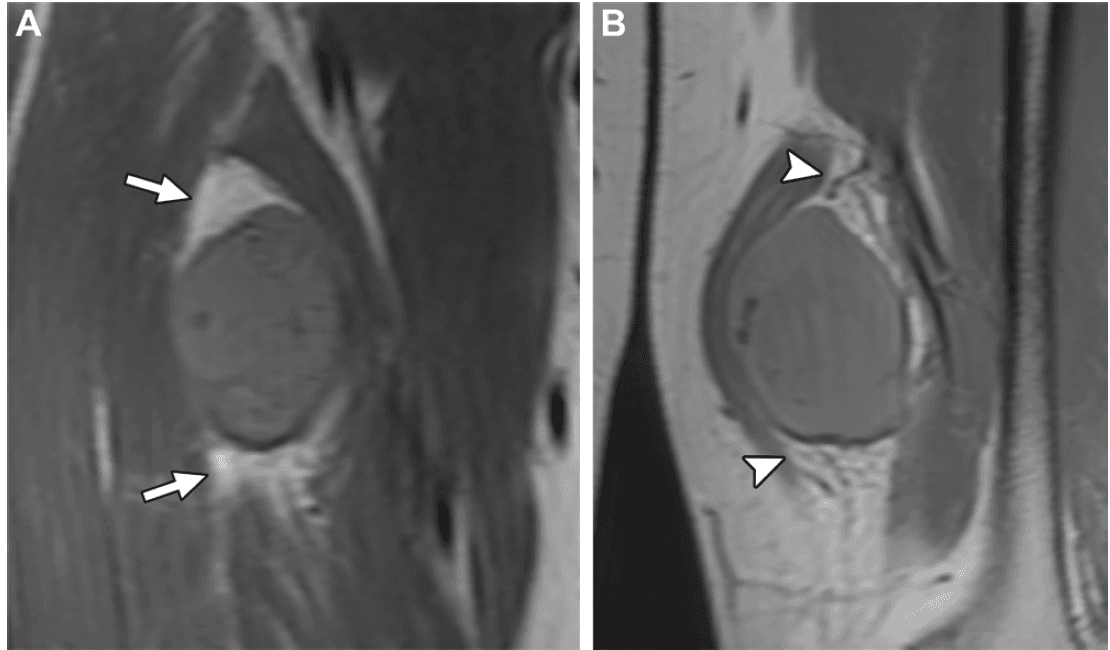
6️⃣ Relationship to Adjacent Structures 🔑 The key factor in limb salvage vs amputation 🧈 Fat plane presence/absence is critical — best seen on axial non–fat-suppressed T1 (always include this sequence!) 🧵 Fascia = natural barrier; subcutaneous malignancies more likely to perforate it or show wide contact
🦴 Bone invasion (uncommon, 5–12%, but worse prognosis when present):
✅ Most accurate sign: abnormal cortical/medullary signal
⚠️ Bone abutment alone (no signal change) has low specificity for invasion
✅ Normal bone–soft tissue interface + peritumoral edema only → strongly favors no invasion
🧓 Periosteum-only abutment → periosteal resection alone may be an adequate margin, but periosteum thins with age (less reliable in older patients) and periosteal stripping may need bone stabilization
⚠️ Pitfall: benign lesions (e.g., tenosynovial giant cell tumor) can also erode bone
🩸 Neurovascular bundle: ↔️ Displaced vs encased 📐 180° absent fat plane = proposed cutoff for encasement 🚫 >270° involvement → usually can't dissect free without violating the tumor 💉 Vessels → can be resected + reconstructed ⚡ Nerves → often lost if truly encased (rarely invaded, but hard to prove on imaging) ✂️ Small abutment → may allow "peeling" the tumor off (neurolysis), leaving the adventitia/epineurium — still negative margins
7️⃣ Signal Intensity & Enhancement Pattern ⚪ High T1 → fat, blood, or melanin ⚫ Low T2 → flow voids, blood, fibrous tissue, calcification 💧 Fluid signal → cystic material, myxoid tissue, blood, or necrosis — postcontrast images needed to sort these out 🚫 Necrotic areas → avoid sampling here for biopsy (inadequate tissue), but don't avoid all necrosis — it's a grading factor for some tumors
🎯 DWI (part of standard protocol): 📈 Higher grade → usually more restriction 🎯 Helps target the highest-cellularity biopsy spot (↑ DWI / ↓ ADC) ⚠️ Pitfall: benign tenosynovial giant cell tumor can also show restriction ⚠️ Pitfall: malignant chondroid/myxoid tumors may show less restriction
🚨 Critical caveat: signal intensity and enhancement pattern cannot reliably separate benign from malignant, or reliably determine histologic grade — useful for narrowing the differential only
8️⃣ Peritumoral Tail, Edema & Enhancement 〰️ Tail sign = thick fascial enhancement extending from the tumor margin 🌫️ Peritumoral edema = ↑T2 signal, feathery/infiltrative borders, no mass effect 🎨 Peritumoral enhancement = enhances beyond the tumor border, also no mass effect 👉 Enhancement (rather than edema alone) is more likely to represent tumor-invaded tissue ⚠️ Benign and intermediate-grade lesions can show tails too: myxoma, nodular fasciitis, fibromatosis 🎯 Matters for how much surrounding tissue to resect + radiation field size
9️⃣ Vasculature 🩸 Feeding/draining vessels — intratumoral or peritumoral — matter for anticipating ligation at surgery 🌀 Flow voids → classic for vascular malformation, alveolar soft-part sarcoma, solitary fibrous tumor 📈 Peritumoral flow voids can also signal a higher tumor grade 🧊 Tumor thrombus (filling defect in a vessel) is rare but important — needs resection + reconstruction 🩸 Leiomyosarcoma can arise directly from a vein
🔟 Lymph Nodes 📊 Uncommon in extremity STS (1.6–12%) 🎯 Higher-risk subtypes: clear cell sarcoma, rhabdomyosarcoma, angiosarcoma, epithelioid sarcoma 🤔 Synovial sarcoma — traditionally included, but recent data has questioned this
Clinical use note
This content is educational and is not patient-specific medical advice. Every phrase, template, classification result, and recommendation must be verified and adapted by a physician using the complete examination, clinical context, current guidelines, and institutional protocol.
Apply the system with a ready-to-edit template
Open an examination-specific framework in RadPhrases, adapt it to the findings, and verify it before signing.