
Knee MRI Report Template: Normal Example and Reporting Checklist
Use a complete normal knee MRI report, a compartment-by-compartment checklist, and careful wording for meniscal and ligament findings.
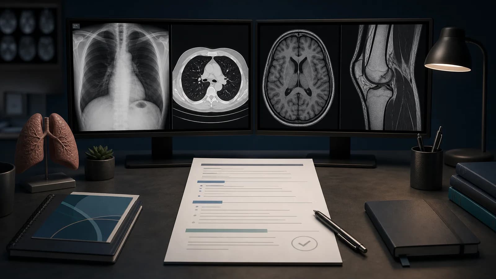
Normal knee MRI report: complete example
This framework is for a routine, noncontrast MRI of one knee. Confirm the side, protocol, image coverage, and clinical question before using it. A normal template is appropriate only after the acquired images have been reviewed; it does not establish that disease outside the protocol or field of view is absent.
Examination: MRI of the right knee without intravenous contrast.

Clinical indication: [Indication.]

Comparison: [Prior study and date / None available.]

Technique: Multiplanar, multisequence MRI of the right knee was performed without intravenous contrast.

Findings: The osseous alignment is maintained. Marrow signal is within expected limits, with no acute fracture or focal osteochondral defect identified. The articular cartilage of the medial and lateral femorotibial compartments and the patellofemoral compartment is preserved. The medial and lateral menisci are normal in morphology and signal, without a surfacing tear or displaced fragment. The anterior and posterior cruciate ligaments and the medial and lateral supporting ligament complexes are intact. The extensor mechanism is intact. No significant joint effusion or popliteal cyst is present. The visualized muscles and soft tissues are unremarkable.

Impression: No significant abnormality identified on this routine MRI of the right knee.Web Report Writer requires an account and Premium access.
Browse all radiology report templates →Adapt the wording to the actual sequences and coverage. If a finding is present, remove the conflicting normal sentence and describe the finding precisely.
Before opening the images
- Match the side on the order, images, and report. Check for a laterality marker and any discrepancy in the supplied clinical history.
- Confirm whether the examination is routine noncontrast MRI, arthrography, or a tailored postoperative, inflammatory, mass, or infection study.
- Review the whole acquired volume, not only the structure named in the indication. Confirm that the meniscal roots, posterior corners, patellofemoral joint, and extensor mechanism are included.
- Check motion, susceptibility, incomplete coverage, and other artifacts. Name a limitation only when it affects evaluation.
- Identify the most relevant prior radiographs, MRI, or operative history. A postoperative meniscus requires comparison with the procedure and prior images when available.
Compartment-by-compartment review
Bones, cartilage, and alignment
Review distal femur, proximal tibia and fibula, patella, and included marrow. Look for fracture, contusion, focal marrow lesion, osteochondral abnormality, and subchondral change. Assess the medial and lateral femorotibial cartilage separately from the patellofemoral cartilage. If cartilage loss is present, state its location and depth where confidently assessed; do not label an MRI appearance alone as clinical osteoarthritis.
Menisci
Trace the medial and lateral menisci through the anterior horn, body, posterior horn, and roots. Assess morphology, displaced tissue, extrusion, and signal reaching an articular surface. If a tear is present, describe the side, segment, surface, pattern when clear, and any displaced fragment or root involvement. Avoid inferring acuity, cause, stability, or repairability from MRI alone. A routine MRI cannot establish the mechanical stability of a small tear, and a meniscal root tear may not always be excluded.
Cruciate and collateral ligaments
Assess ACL and PCL continuity, course, signal, and secondary signs of insufficiency. Review the superficial and deep MCL, fibular collateral ligament, popliteus tendon, and other posterolateral supporting structures as indicated. Describe a ligament injury as partial- or full-thickness only when supported by the images; partial tears can be difficult to characterize. Check for adjacent edema, avulsion, and alignment changes.
Extensor mechanism and supporting tissues
Review quadriceps and patellar tendons, patella, retinacula, and medial patellofemoral ligament when clinically relevant. Inspect the joint recesses, bursae, popliteal region, muscles, and visible soft tissues. A small amount of fluid or nonspecific edema should not be upgraded to a diagnosis without supporting morphology and clinical context.
Focused wording modules
Use these only when they match the images:
Meniscus — normal: The medial and lateral menisci are normal in morphology and signal, without a surfacing tear or displaced fragment.

Meniscus — tear: A [horizontal / vertical-longitudinal / radial / complex] tear involves the [anterior horn / body / posterior horn / root] of the [medial / lateral] meniscus, extending to the [superior / inferior / both] articular surfaces. [No displaced fragment is identified.]

Meniscus — equivocal: Intrasubstance signal in the [segment] of the [medial / lateral] meniscus approaches but does not clearly reach an articular surface. No displaced fragment is seen.

Ligament — sprain: The [ligament] is mildly thickened and increased in signal with fibers remaining in continuity, compatible with a low-grade sprain. No avulsion is identified.

Ligament — tear: Full-thickness discontinuity of the [ligament] is present at the [site], with [associated edema / avulsion / secondary alignment finding].
Do not use “grade 1, 2, or 3” as a substitute for describing what is visible unless a locally adopted system specifically requires it. For a bone-contusion pattern and associated injury review, see Bone Contusion Patterns of the Knee.
Fictional worked example: twisting injury
The following teaching case is fictional. It shows how the same complete report structure can be adapted when the examination is abnormal. The report does not infer when the tear occurred or recommend treatment.
Examination: MRI of the right knee without intravenous contrast.

Clinical indication: Twisting injury with knee pain.

Comparison: Right knee radiographs dated [date].

Technique: Multiplanar, multisequence MRI of the right knee was performed without intravenous contrast.

Findings: There is a complete midsubstance tear of the anterior cruciate ligament with surrounding edema. Bone marrow edema-like signal is present in the posterolateral tibial plateau and lateral femoral condyle. No fracture line or displaced osteochondral fragment is identified. The posterior cruciate ligament and collateral ligament complexes are intact. The medial and lateral menisci are normal in morphology, without a surfacing tear or displaced fragment. The articular cartilage is maintained. There is a small joint effusion. The extensor mechanism is intact.

Impression:
1. Complete midsubstance anterior cruciate ligament tear.
2. Contusions of the posterolateral tibial plateau and lateral femoral condyle. No fracture or displaced osteochondral fragment.
The first impression item names the principal injury; the second records the osseous footprint and its relevant negative findings. Do not describe marrow edema as a fracture unless a fracture line or other convincing evidence is present.
Protocol limits and wording pitfalls
Routine knee MRI usually includes multiplanar fluid-sensitive and T1-weighted information, but local protocols differ. The ESSR recommendations support review in three orthogonal planes and assessment beyond the named indication. If the requested question requires different coverage or sequences, report what was and was not evaluated rather than implying a comprehensive examination.
Avoid these shortcuts:
- “No internal derangement” when the menisci, ligaments, cartilage, and extensor mechanism have not all been assessed.
- Calling a tear acute, chronic, unstable, repairable, or the cause of pain without adequate evidence.
- Calling nonspecific intrameniscal signal a tear when it does not reach a surface and morphology is preserved.
- Calling mild cartilage change “osteoarthritis” without clinical and radiographic context.
- Using a normal nonoperative template for a postoperative knee without accounting for altered anatomy and comparison studies.
- Reporting an unqualified negative result when motion or limited coverage obscures a structure.
Primary references
- Parkar AP, Adriaensen MEAP, et al. ESR Essentials: MRI of the knee—practice recommendations by ESSR. European Radiology. 2024;34:6590–6599. doi:10.1007/s00330-024-10706-7 (opens in a new tab)
- American College of Radiology, Society for Pediatric Radiology, Society of Skeletal Radiology. Practice Parameter for the Performance and Interpretation of MRI of the Knee. Revised 2025, Resolution 7. Official practice parameter (opens in a new tab)
- Nguyen JC, Boutin RD, et al. Standardized MRI nomenclature for knee meniscal lesions: SSR consensus panel report. Skeletal Radiology. 2026. doi:10.1007/s00256-026-05281-5 (opens in a new tab)
This educational framework does not replace patient-specific interpretation, current local protocols, or clinical judgment.
Clinical use note
This content is educational and is not patient-specific medical advice. Every phrase, template, classification result, and recommendation must be verified and adapted by a physician using the complete examination, clinical context, current guidelines, and institutional protocol.
Use a review-first reporting workflow
Download RadPhrases for its curated phrase library and text expansion. Check every inserted phrase against the images and clinical question before signing.
Related reporting resources
Browse the other checklists, phrase sets, and systematic approaches in this topic.