
Brain MRI Reporting Checklist
A systematic guide to assessing sequences, diffusion, blood products, parenchyma, extra-axial spaces, and adjacent structures on routine adult brain MRI.
RadPhrases Editorial Team6 min de lectura
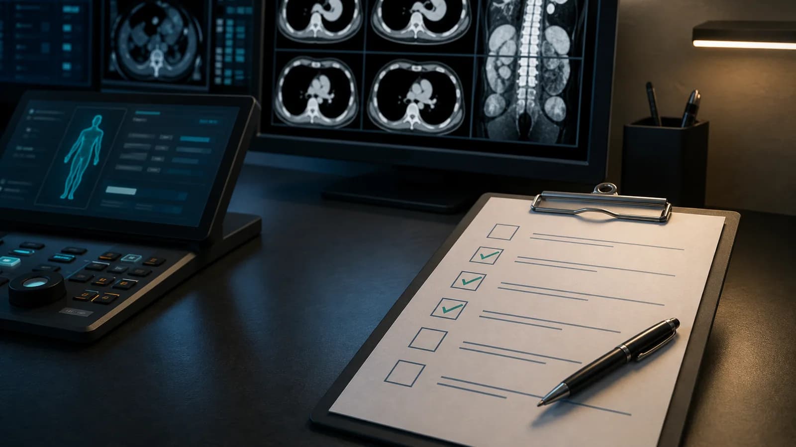
A systematic guide to assessing sequences, diffusion, blood products, parenchyma, extra-axial spaces, and adjacent structures on routine adult brain MRI.
A systematic guide to assessing sequences, diffusion, blood products, parenchyma, extra-axial spaces, and adjacent structures on routine adult brain MRI.
| Scope This content is a general checklist for routine adult brain MRI. Epilepsy, pituitary, internal auditory canal, demyelinating disease, tumor perfusion/spectroscopy, vascular MRI, pediatric, and postoperative examinations require dedicated protocols. |
|---|
Brain MRI includes numerous sequences and contrast mechanisms. The same lesion provides different information on diffusion, FLAIR, susceptibility, and postcontrast images. The examination should therefore be read by cross-checking the same anatomical region across sequences rather than reviewing each sequence in isolation.
This checklist can be used to confirm the adequacy of the basic sequences and inclusion of important anatomical regions in routine adult brain MRI reports.
☐ Read the indication. Headache, seizure, focal deficit, cognitive change, tumor follow-up, and suspected infection place different emphasis on the sequences.
☐ Confirm the protocol. Check for T1, T2, FLAIR, DWI/ADC, and susceptibility sequences and, when contrast was administered, appropriate postcontrast images.
☐ State contrast information clearly. Gadolinium administration, dose information, and the presence of postcontrast sequences should be documented in the technique.
☐ Motion and artifact. Specify which region is limited by susceptibility, dental material, flow, or motion artifact.
☐ Open the prior examination. Consider dates and protocol similarity when assessing lesion burden, size, enhancement, and volume change.
☐ DWI and ADC. Distinguish true restricted diffusion from T2 shine-through and carefully assess the artifact-prone skull base and posterior fossa.
☐ FLAIR/T2. Check for edema, gliosis, white-matter lesions, cortical-subcortical signal abnormality, and CSF suppression.
☐ T1-weighted images. Use them to assess anatomy, fat or blood products, atrophy, and intrinsically T1-hyperintense foci before contrast.
☐ SWI or GRE. Assess microhemorrhage, hemosiderin, cavernous malformation, venous structures, and possible calcification; consider CT correlation when needed.
☐ Postcontrast T1. Systematically survey for parenchymal, leptomeningeal, pachymeningeal, and cranial nerve enhancement.
☐ Additional sequences. When perfusion, spectroscopy, 3D imaging, or angiography is present, assess it separately according to the indication and do not insert assumptions automatically into the standard report template.
☐ Acute ischemia. Determine location and distribution on DWI/ADC and interpret together with FLAIR and vascular signal findings.
☐ Acute or subacute hemorrhage. Assess susceptibility and T1/T2 signal together and state any mass effect or intraventricular extension.
☐ Herniation and hydrocephalus. Check the ventricles, basal cisterns, midline, and foramen magnum level.
☐ Suspected infection/abscess. Assess diffusion, ring enhancement, surrounding edema, and meningeal findings in the clinical context.
☐ Clues to venous thrombosis or major-vessel disease. Distinguish the scope of routine MRI/MRA and consider recommending a dedicated vascular examination when needed.
☐ Cortical signal and thickness. Assess for focal thickening, FLAIR signal change, atrophy, and clues to cortical developmental abnormality.
☐ Subcortical and deep white matter. Describe lesion number, size, distribution, and morphology; avoid an overly definitive diagnosis for nonspecific findings.
☐ Deep gray nuclei. Check symmetry, signal, and diffusion changes in the basal ganglia and thalami.
☐ Corpus callosum. Assess signal, thickness, lesions, and developmental or postoperative changes.
☐ Medial temporal structures. Review hippocampal symmetry and signal according to the indication; when no epilepsy protocol was performed, state the limits of detailed assessment.
☐ Brainstem. Survey the midbrain, pons, and medulla in axial and sagittal planes for signal abnormality, mass, and atrophy.
☐ Cerebellum. Is there ischemia, a mass, atrophy, or developmental change in the hemispheres, vermis, or peduncles?
☐ Fourth ventricle and cisterns. Assess obstruction, mass effect, and visible findings involving the cisternal segments of the cranial nerves.
☐ Craniocervical junction. Check the tonsillar level, foramen magnum, and imaged upper cervical cord.
☐ Ventricular size and symmetry. Distinguish enlargement due to atrophy from hydrocephalus using clinical and morphologic findings.
☐ Transependymal CSF flow. Try to distinguish periventricular FLAIR signal from chronic white-matter change.
☐ Sulci and cisterns. Assess age appropriateness, focal effacement, and extra-axial mass effect.
☐ Subdural/epidural spaces. Cross-check sequences for collections, blood products, or signs of empyema.
☐ Dura and leptomeninges. On contrast-enhanced examinations, state the distribution of thickening and enhancement.
☐ Sella and pituitary. Within the limits of routine sections, note an obvious mass or empty-sella appearance; a dedicated protocol may be required for microadenoma assessment.
☐ Orbits and optic pathways. Assess an obvious mass, signal abnormality, or asymmetry within image-quality limitations.
☐ Paranasal sinuses. Note fluid levels, mucosal thickening, and mass-like opacification in the clinical context.
☐ Mastoid air cells. Check for fluid/opacification and signs of adjacent complications.
☐ Skull and scalp. Assess marrow signal, masses, and soft-tissue lesions to the extent included in the field of view.
| Situations requiring urgent communication on MRI For acute ischemia, acute hemorrhage, herniation, obstructive hydrocephalus, abscess, or other findings requiring rapid management, the final report alone may not be sufficient. The clinical team should be informed within the time and by the method defined by the institution, and the communication should be documented in the report or system. |
|---|
| INDICATION: [Clinical question] TECHNIQUE: Multiplanar, multisequence brain MRI. [IV contrast administered/not administered]. [Sequences and limitations]. COMPARISON: Examination dated [date]. FINDINGS: - Diffusion: [...] - Blood products/susceptibility: [...] - Cerebral parenchyma and white matter: [...] - Deep gray nuclei / brainstem / cerebellum: [...] - Ventricles, cisterns, and mass effect: [...] - Extra-axial spaces and meninges: [...] - Sella/orbits/sinuses/mastoids/craniocervical junction: [...] - Enhancement: [if applicable] IMPRESSION: 1. [Primary clinical finding.] 2. [Interval change / secondary finding.] 3. [Dedicated protocol or follow-up recommendation, if needed.] |
|---|
Este contenido es educativo y no constituye asesoramiento médico específico para un paciente. Antes de firmar, el médico debe verificar y adaptar cada frase, plantilla, resultado de clasificación y recomendación teniendo en cuenta todas las imágenes, el contexto clínico, las guías vigentes y el protocolo del centro.
Use this sequence as an editable RadPhrases template and verify it against the images in every case.
Explora otras listas de comprobación, expresiones y métodos sistemáticos de este tema.