
Chest CT Reporting Checklist
A systematic approach to comprehensively reviewing the lung parenchyma, airways, pleura, mediastinum, cardiovascular structures, and upper abdomen on routine diagnostic chest CT.
RadPhrases Editorial Team5 min de lectura
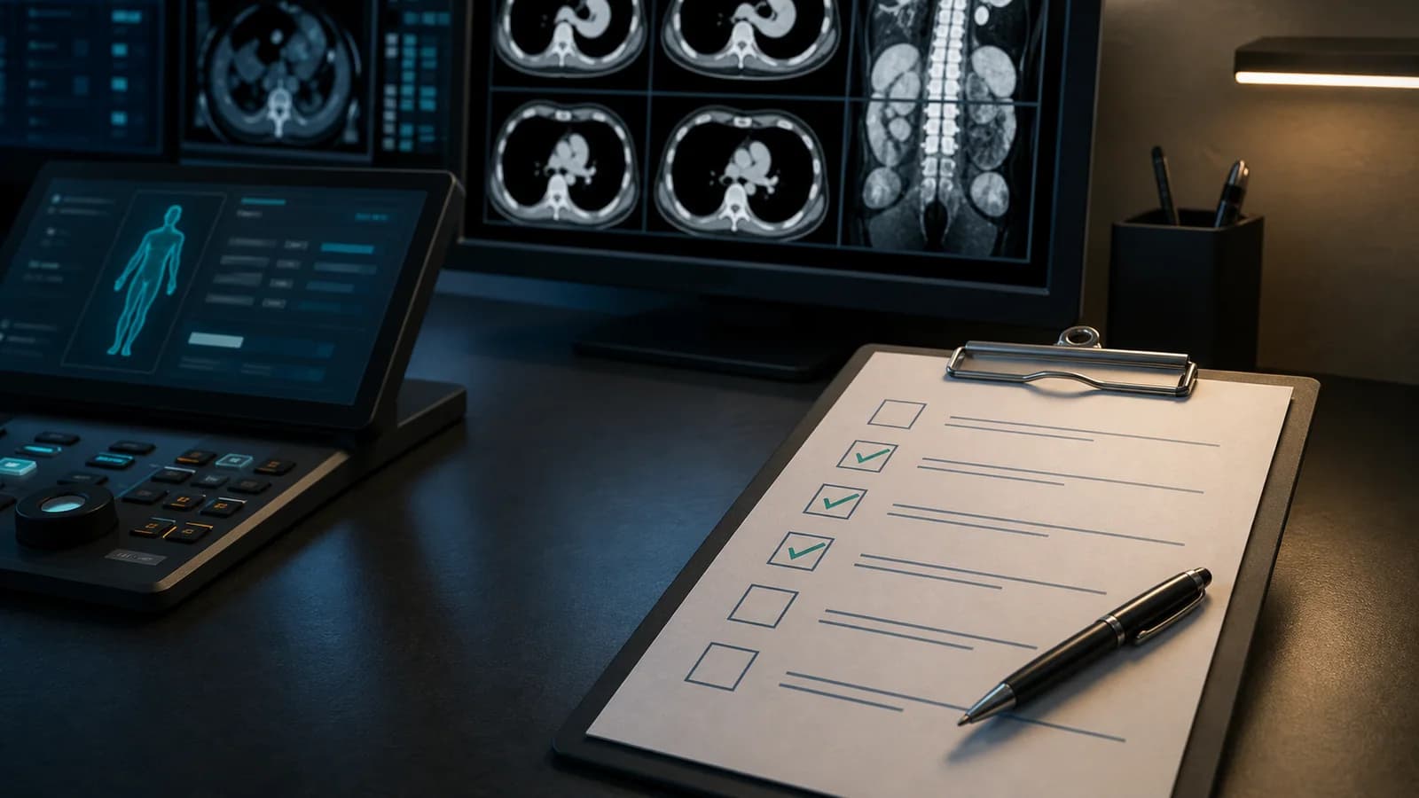
A systematic approach to comprehensively reviewing the lung parenchyma, airways, pleura, mediastinum, cardiovascular structures, and upper abdomen on routine diagnostic chest CT.
A systematic approach to comprehensively reviewing the lung parenchyma, airways, pleura, mediastinum, cardiovascular structures, and upper abdomen on routine diagnostic chest CT.
| Scope This content provides a general reading sequence for routine diagnostic chest CT. CT pulmonary angiography, aortic CT angiography, high-resolution CT, lung cancer screening, and oncologic response assessment require separate protocols and reporting systems. |
|---|
Chest CT is not a single-organ examination. In addition to the lung parenchyma, the airways, pleura, mediastinum, lymph nodes, cardiovascular structures, chest wall, and imaged upper abdomen are all part of the report.
A systematic reading sequence helps prevent mediastinal or upper abdominal findings from being overlooked while attention is concentrated on the lungs. This draft provides a repeatable checklist for routine adult chest CT reporting.
☐ Read the indication. Infection, malignancy, nodule follow-up, interstitial disease, trauma, and postoperative assessment require different priorities.
☐ Identify contrast status and phase. Noncontrast, venous phase, or a dedicated angiographic phase determines the limits of vascular and mediastinal interpretation.
☐ Assess inspiration and motion. Poor inspiration may increase dependent opacities and create a false mosaic appearance.
☐ Confirm coverage. The entire thorax from the apices to the costophrenic angles, together with the required upper abdomen, should be included.
☐ Open the prior examination. For changes in nodules, masses, lymph nodes, and pleural fluid, note the date and similarity of technique.
☐ Pneumothorax and large pleural collection. Is there mediastinal shift or cardiac compression suggesting tension physiology?
☐ Obvious aortic or vascular emergency. Within the limits of a routine protocol, do not miss visible signs such as a dissection flap, rupture, or active bleeding.
☐ Large-airway obstruction. Is there a foreign body, mucus plug, or tumor-related obstruction in a main bronchus?
☐ Extensive acute parenchymal process. Dense consolidation, edema, or severe aspiration may alter clinical priority.
☐ Device complication. Quickly check tubes, catheters, drains, and surgical material.
☐ Use a fixed lobar and segmental sequence. Review both lungs from apices to bases on axial images and confirm findings on coronal and sagittal reconstructions.
☐ Nodule and mass. Describe location, size, morphology, margins, attenuation, and change from prior imaging.
☐ Consolidation and ground-glass opacity. Assess distribution, lobular or segmental pattern, air bronchograms, and associated volume loss.
☐ Atelectasis. Interpret possible passive, obstructive, or scar-related appearances together with bronchial patency and pleural disease.
☐ Interstitial findings. Note reticulation, septal thickening, traction bronchiectasis, honeycombing, and distribution; state when routine CT may not substitute for HRCT.
☐ Emphysema and clues to air trapping. Assess type, distribution, and associated bullae; if no expiratory series is available, remain cautious about air-trapping assessment.
☐ Cavity/cyst. Describe wall thickness, contents, surrounding parenchyma, number, and distribution.
☐ Trachea and main bronchi. Assess the lumen, wall, secretions, and extrinsic compression.
☐ Bronchiectasis. Review the bronchoarterial ratio, visibility of peripheral bronchi, wall thickening, and mucus plugging.
☐ Endobronchial lesion. Check for focal narrowing, cutoff, or associated distal atelectasis/consolidation.
☐ Suspected tracheobronchomalacia. State the limitations of a single-phase inspiratory examination; dynamic assessment may be required.
☐ Pleural fluid. Assess volume, side, loculation, pleural thickening, and adjacent atelectasis.
☐ Pneumothorax. Check for small anterior or basal air collections on multiplanar images.
☐ Pleural nodule/plaque. Note calcification, extent, and suspected invasion.
☐ Diaphragm. Is there elevation, hernia, defect, or focal contour abnormality?
☐ Mediastinal compartments. Systematically assess for masses, fluid, air, and distortion of fat planes.
☐ Lymph nodes. State station, short-axis size, morphology, and change from prior imaging, interpreted in the clinical context.
☐ Hila. Review the vessels, lymph nodes, and bronchi together and try to explain any asymmetry.
☐ Esophagus. Is there marked wall thickening, dilatation, hiatal hernia, or alteration of the surrounding fat planes?
☐ Thymus and visible thyroid. Note an obvious mass or nodular enlargement according to age and clinical context.
☐ Cardiac size and chambers. Assess obvious enlargement while respecting the measurement limitations of routine nongated CT.
☐ Pericardium. Is there fluid, thickening, or calcification?
☐ Aorta. Assess caliber, atherosclerosis, and visible acute abnormality; when measurements are needed, pay attention to the imaging plane.
☐ Pulmonary arteries. Check the main pulmonary artery caliber and visible filling defects; when appropriate, state that embolism cannot be excluded on a nondedicated examination.
☐ Coronary calcification. Report conspicuous calcification in keeping with image quality, remembering that this is not a substitute for cardiac CT.
☐ Ribs, sternum, vertebrae, and shoulder girdle. Review bone windows for fracture, destruction, sclerotic foci, or compression deformity.
☐ Soft tissues. Is there an obvious mass or collection in the breasts, axillae, subcutaneous tissues, or muscle planes?
☐ Upper abdomen. Systematically assess the imaged portions of the liver, adrenal glands, spleen, pancreas, and kidneys.
☐ Surgical and radiotherapy changes. Compare with prior images to distinguish expected changes from complications or suspected recurrence.
| Emergency findings can occur on routine CT Tension pneumothorax, large-airway obstruction, massive hemothorax, suspected aortic rupture/dissection, substantial device malposition, or other urgent findings should trigger the local critical results communication process. If uncertainty results from a nondedicated protocol, clearly communicate that limitation to the clinical team. |
|---|
| INDICATION: [Clinical question] TECHNIQUE: [Noncontrast / IV contrast-enhanced] chest CT. [Phase, reconstructions, and limitations]. COMPARISON: Examination dated [date]. FINDINGS: - Lung parenchyma: [...] - Airways: [...] - Pleura: [...] - Mediastinum/hila/lymph nodes: [...] - Heart/pericardium/great vessels: [...] - Chest wall and bones: [...] - Imaged upper abdomen: [...] IMPRESSION: 1. [Primary clinical finding.] 2. [Interval change / important secondary finding.] 3. [Context-based recommendation for follow-up or further evaluation, when appropriate.] |
|---|
Este contenido es educativo y no constituye asesoramiento médico específico para un paciente. Antes de firmar, el médico debe verificar y adaptar cada frase, plantilla, resultado de clasificación y recomendación teniendo en cuenta todas las imágenes, el contexto clínico, las guías vigentes y el protocolo del centro.
Use this sequence as an editable RadPhrases template and verify it against the images in every case.
Explora otras listas de comprobación, expresiones y métodos sistemáticos de este tema.