
Common Pelvic CT Reporting Phrases
Use measured pelvic CT wording for bladder, pelvic organs, bowel, fluid, lymph nodes, and bone findings.
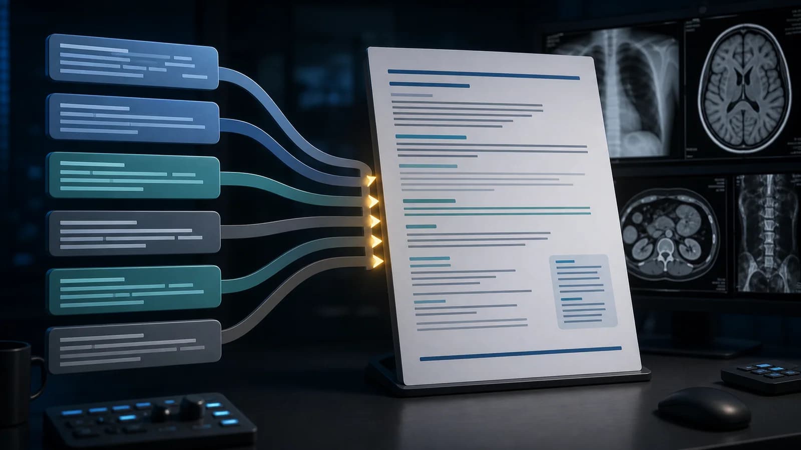
Pelvic CT wording should reflect the structure actually seen, the contrast phase, and technical limits. An underfilled bladder, unopacified vessel, collapsed bowel loop, or cycle-related ovarian appearance should not be converted into a definite disease statement. A stock phrase belongs in the report only after image confirmation.
These are teaching examples for adult pelvic CT. Remove irrelevant lines according to sex, surgical history, contrast phase, oral contrast, and clinical indication. An acute critical finding requires direct communication.
Assessment framework
- Bladder: The bladder is adequately distended without a conspicuous focal wall mass.
- Bowel: No obstruction or free air is identified in the pelvic bowel loops.
- Fluid: A small amount of pelvic free fluid is present and should be interpreted in context.
- Nodes: No enlarged pelvic node with suspicious morphology is identified.
- Bone: No acute fracture or aggressive lytic lesion is seen in the included osseous structures.
What the report should contain
- Use uterus or prostate wording only for the correct patient.
- For an adnexal lesion, state side, size, solid tissue, and signs of acute complication.
- Describe loss of a fat plane as suspicious contact rather than automatic invasion.
- Connect a distal ureteric finding with hydronephrosis in the same impression.
- Preserve CT limits for superficial mucosal disease and small endometriosis implants.
Common errors
- Calling an underfilled bladder definitively thick walled.
- Treating a physiologic ovarian follicle as neoplasm.
- Using a normal organ list in place of the clinical answer.
Example report wording
Example impression: A 34 mm thin-walled homogeneous fluid-attenuation cystic lesion is present in the left adnexa, without a solid nodule or surrounding inflammation. There is a small amount of pelvic free fluid. CT shows no acute sign of torsion, but persistent clinical concern for torsion requires ultrasound and clinical assessment.
Use the systematic abdomen and pelvis CT reporting guide for the complete examination.
References
- American College of Radiology. Practice Parameter for MRI of the Soft-Tissue Components of the Pelvis. Source (opens in a new tab)
- American College of Radiology. Practice Parameter for CT of the Abdomen and Pelvis. Source (opens in a new tab)
Clinical note: This material is for radiology education. Do not generate an automatic diagnosis or management decision without checking guideline scope, patient factors, and local policy.
Clinical use note
This content is educational and is not patient-specific medical advice. Every phrase, template, classification result, and recommendation must be verified and adapted by a physician using the complete examination, clinical context, current guidelines, and institutional protocol.
Adapt the report framework to the case
Use this review order with an editable RadPhrases template and verify every line against the images.
Related calculators
Browse the other checklists, phrase sets, and systematic approaches in this topic.