
Incidental Solid Pulmonary Nodule Reporting: Measurement and Impression Examples
A practical framework for measuring incidental solid lung nodules, documenting prior comparison, and applying Fleischner 2017 recommendations.
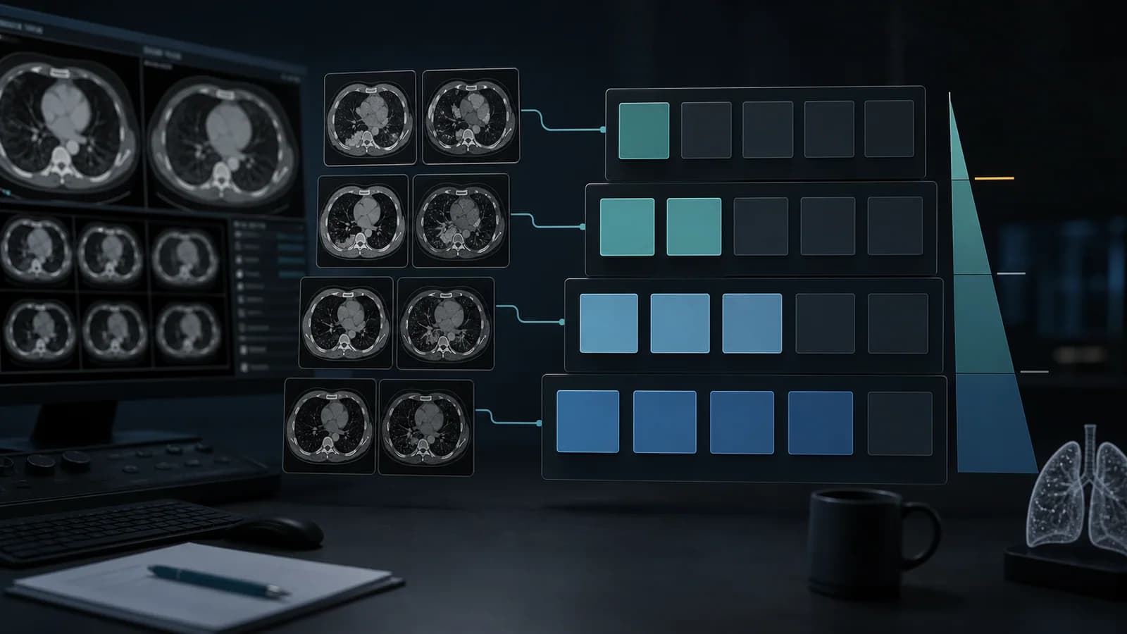
Incidental solid nodule report framework
Use this structure for an incidentally detected solid pulmonary nodule on CT. Complete the bracketed fields, document the comparison, and make any follow-up statement only after checking that the patient and examination fit the cited pathway.
Examination: CT of the chest [without/with] intravenous contrast. [Thin-section lung reconstructions available.]

Comparison: [Prior chest CT dated DD Month YYYY / No prior chest CT available.]

Findings: A [solid, noncalcified] pulmonary nodule is present in the [right/left] [upper/middle/lower] lobe, [segment/location], measuring [long axis] × [short axis] mm (mean diameter [rounded] mm) on [series/image]. Margins are [smooth/lobulated/spiculated/other]. [Describe macroscopic fat or calcification if present.] [No additional pulmonary nodules / Additional nodules are listed below.] [Relevant background risk features, such as emphysema or pulmonary fibrosis, are described.]

Impression:
1. [Location and morphology] solid pulmonary nodule measuring [mean] mm, [unchanged from / new since / not previously imaged]. [For an eligible incidental nodule, state the selected guideline-based option and interval, if appropriate.]
2. [Additional clinically relevant finding, if any.]Web Report Writer requires an account and Premium access.
Browse all radiology report templates →This is a reporting framework, not a patient-specific management decision. Do not complete a negative statement unless the relevant images and structures were reviewed. If the nodule is calcified, fat-containing, part-solid, or ground-glass, describe that accurately and use the guidance intended for that finding.
Confirm the pathway before applying a size threshold
The Fleischner Society 2017 recommendations address incidentally detected nodules in adults. They are not intended for patients younger than 35 years, patients with known cancer at risk for pulmonary metastases, or immunocompromised patients in whom infection is a concern. They are not a screening system: nodules detected on a lung cancer screening examination should be assessed with the applicable ACR Lung-RADS release. Subsolid nodules have separate recommendations.
Record enough context to explain why a recommendation was selected:
- Was the nodule found incidentally on a diagnostic CT, rather than on a screening examination?
- Is the patient within the guideline's intended population?
- Is the lesion truly solid, and is it a nodule rather than scar, vessel, atelectasis, or a typical perifissural intrapulmonary lymph node?
- Is there a prior CT that can establish stability or growth?
- Are age, smoking exposure, upper-lobe location, suspicious morphology, emphysema, or pulmonary fibrosis relevant to estimated risk?
- Is the CT technically comparable with the prior examination?
Risk is not determined by smoking history alone. The recommendation ranges leave room for morphology, patient risk, comorbidity, and preferences; explain a deliberate departure from a size-based interval when clinically relevant.
Measure consistently and make the comparison reproducible
Thin contiguous sections, generally 1.5 mm or less, help characterize and measure small nodules. On the image plane that shows the greatest dimensions, measure the long and short axes on the same image, average them, and record the result to the nearest whole millimetre. Avoid decimal precision that the method cannot support. For nodules larger than 10 mm, reporting both axes in addition to the mean can be useful.
When a prior CT is available, compare the same nodule directly using similar planes, reconstructions, and lung windows. Record the prior date and the earlier measurement if known. A small difference can reflect technique or measurement variability; do not label growth from a few tenths of a millimetre or from mismatched slice thickness alone. If the baseline study has thick sections or incomplete coverage, state that limitation.
Measurement worksheet
| Field | Record |
|---|---|
| Nodule | [Index nodule; if multiple, identify each relevant lesion] |
| Location | [Side, lobe, segment, central/peripheral or perifissural] |
| Attenuation and morphology | [Solid; calcification/fat; margin; shape] |
| Current size | [Long × short axis; mean rounded to whole mm] |
| Image reference | [Series and image; plane/window] |
| Comparison | [Prior date, prior size, stable/growing/new/indeterminate] |
| Context | [Screening or incidental; cancer/immunosuppression; relevant risk factors] |
| Recommendation | [Guideline pathway and chosen next step, when applicable] |
Interpreting common solid-nodule size groups
For a solitary solid nodule smaller than 6 mm, routine follow-up is generally not recommended; a 12-month CT may be considered in a higher-risk patient. For a solitary 6–8 mm nodule, CT at 6–12 months is recommended. A further CT at 18–24 months is recommended for a high-risk patient and may be considered for a low-risk patient, taking morphology, stability, and patient preference into account. For a solitary nodule larger than 8 mm, options include short-interval CT, PET/CT, tissue sampling, or a combination; selection depends on size, morphology, patient factors, and local practice.
Multiple solid nodules have a separate pathway. When all are smaller than 6 mm, routine follow-up is generally not recommended, though a 12-month CT may be considered in a higher-risk patient. If at least one is 6 mm or larger, CT at about 3–6 months is recommended. For multiple nodules in the 6–8 mm group, a further CT at 18–24 months is recommended for a high-risk patient and may be considered for a low-risk patient. If a larger or more suspicious nodule is present, use that lesion to guide management and apply the solitary-nodule recommendations as appropriate. The most suspicious nodule, which may not be the largest, guides the recommendation. Record the other relevant nodules so they can be compared as well.
These intervals summarize the 2017 Fleischner recommendations for adults with incidental nodules; they do not replace the full guideline or patient-specific decision making.
Fictional worked cases
Single 7 mm nodule with no prior CT
Case: A fictional 64-year-old former smoker has a smooth, solid 7 × 6 mm nodule in the apical segment of the right upper lobe on thin-section diagnostic CT. Mild emphysema is also present. No prior CT is available, and there is no known malignancy or immunosuppression. This example uses the higher-risk pathway because of the combined former-smoking history, emphysema, and upper-lobe location; actual risk assessment remains patient-specific.
Examination: Noncontrast CT of the chest with contiguous thin-section lung reconstructions.

Comparison: No prior chest CT available.

Findings: A smooth solid nodule in the apical segment of the right upper lobe measures 7 × 6 mm (mean diameter 7 mm). Mild centrilobular emphysema is present. No additional pulmonary nodule is identified.

Impression: Solitary 7 mm solid right upper lobe nodule. Given this example's higher-risk context, chest CT at 6–12 months and again at 18–24 months is recommended.
The case is fictional. A real report should reflect the patient's risk profile, image quality, morphology, prior studies, and local follow-up practice.
Multiple nodules with one lesion at least 6 mm
Case: A fictional 68-year-old current smoker with mild emphysema has three solid nodules measuring 3 mm, 5 mm, and 7 mm. The 7 mm nodule is mildly lobulated and is in the left upper lobe. There is no prior CT, known cancer, or immunosuppression. The example uses the higher-risk pathway.
Findings: Three solid pulmonary nodules are present: a mildly lobulated 7 mm nodule in the left upper lobe, a smooth 5 mm right lower lobe nodule, and a smooth 3 mm left lower lobe nodule. Mild centrilobular emphysema is present. No prior CT is available for comparison.

Impression: Multiple solid pulmonary nodules, including a 7 mm mildly lobulated left upper lobe nodule. For this higher-risk incidental-nodule scenario, chest CT at approximately 3–6 months and again at 18–24 months is recommended. The left upper lobe nodule is the most suspicious lesion for comparison.
Nodule larger than 8 mm
Case: A fictional 70-year-old has a new 11 mm spiculated solid nodule. The impression can identify the finding and outline options without presenting one as automatic:
Impression: New 11 mm spiculated solid nodule in the right upper lobe. Prompt clinical assessment is advised. Depending on estimated malignancy risk, morphology, comorbidity, and patient preference, options include short-interval chest CT, PET/CT, and/or tissue sampling.Reporting checklist
- State the examination and whether contrast was administered.
- Confirm incidental-nodule versus screening context and guideline eligibility.
- Identify each relevant nodule by side, lobe, and location.
- Describe solid attenuation and relevant morphology.
- Include long and short axes, mean diameter rounded to whole millimetres, and image reference.
- Compare with the most relevant prior CT and state limitations in comparability.
- For multiple nodules, identify the most suspicious lesion and record other lesions for follow-up.
- Keep the impression concise: finding, comparison, and a reasoned next-step statement when appropriate.
Wording pitfalls and limits
Do not use Lung-RADS categories for an incidental diagnostic CT, or apply Fleischner intervals to a screening examination. Do not apply the solid-nodule table to ground-glass or part-solid nodules. Do not automatically describe a nodule as “stable” from a chest radiograph or a technically different partial scan when CT comparison is inadequate. A typical perifissural intrapulmonary lymph node may not need follow-up, but the morphology must actually support that description.
A follow-up recommendation does not diagnose malignancy and does not ensure that follow-up occurs. State the finding and suggested interval clearly, and use the local process for communicating actionable results. CT morphology and interval size change must be interpreted with the complete clinical context.
References
- MacMahon H, Naidich DP, Goo JM, et al. Guidelines for Management of Incidental Pulmonary Nodules Detected on CT Images: From the Fleischner Society 2017. Radiology. 2017;284:228–243. doi:10.1148/radiol.2017161659 (opens in a new tab).
- American College of Radiology. Lung CT Screening Reporting & Data System (Lung-RADS), current release and archive (opens in a new tab).
Clinical note: Educational examples only. Apply current guidance to the actual patient, examination, and local protocol.
Clinical use note
This content is educational and is not patient-specific medical advice. Every phrase, template, classification result, and recommendation must be verified and adapted by a physician using the complete examination, clinical context, current guidelines, and institutional protocol.
Start from an editable report structure
Open a report framework in RadPhrases, then verify each statement against the images, protocol, and clinical context.
Related reporting resources
Browse the other checklists, phrase sets, and systematic approaches in this topic.