
RECIST 1.1 Follow-up CT Reporting: Lesion Tables and Worked Examples
A RECIST 1.1 worksheet for baseline and follow-up CT, target and non-target lesions, new findings, sum calculations, and response wording.
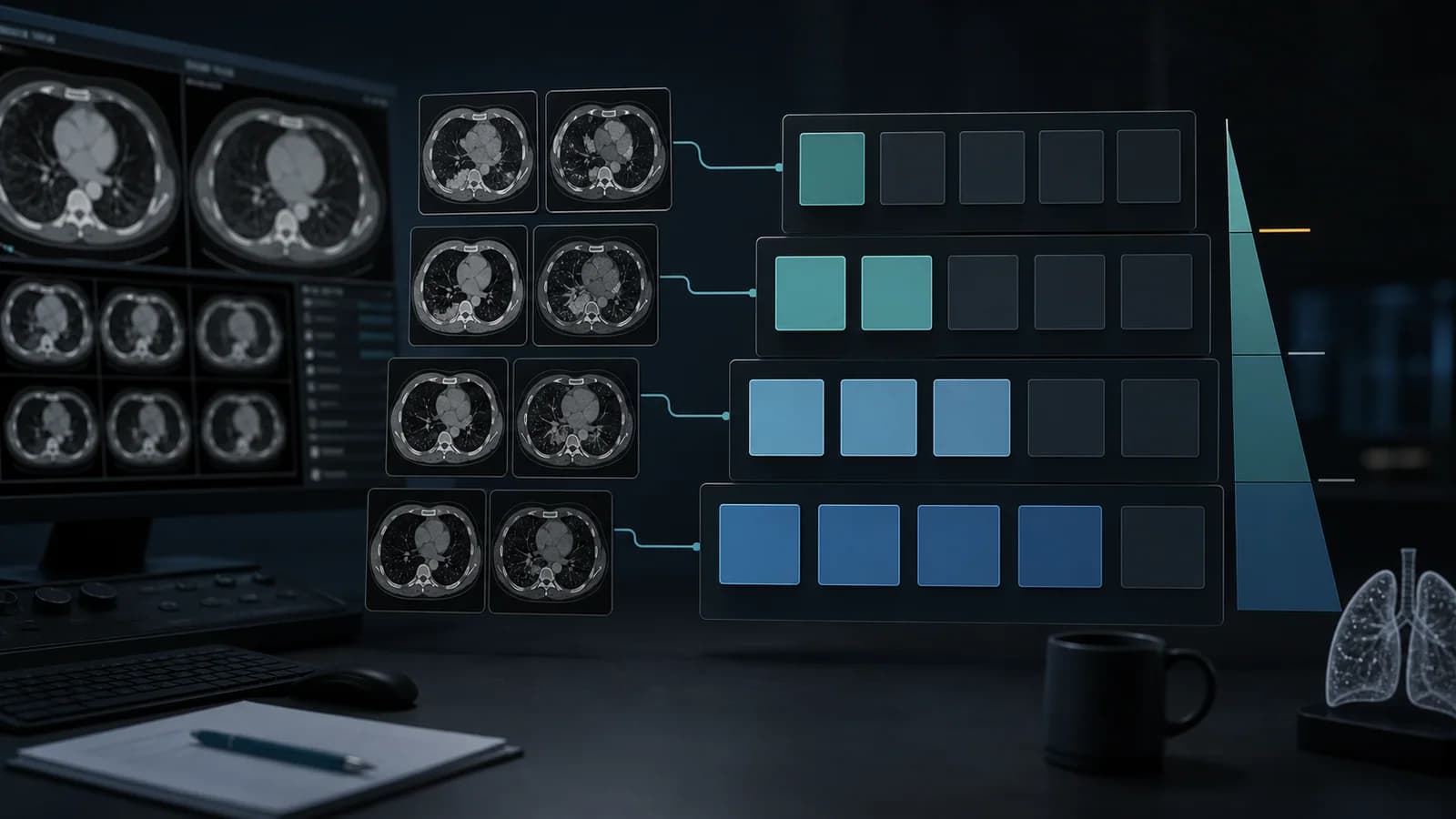
RECIST 1.1 follow-up CT report framework
Use this worksheet when formal response assessment by RECIST version 1.1 is requested and the study protocol uses that version. Confirm the applicable assessment method before measuring: a trial charter or disease-specific protocol may add instructions or use another response system.
Examination: Contrast-enhanced CT of the chest, abdomen, and pelvis. [Specify phases, coverage, and any technical limitation.]

Comparison: RECIST baseline CT dated [date]; most recent CT dated [date].

Target lesions:
1. [Organ/site, series/image]: baseline [ ] mm; nadir [ ] mm; current [ ] mm.
2. [Organ/site, series/image]: baseline [ ] mm; nadir [ ] mm; current [ ] mm.
3. [Add only if selected; maximum five target lesions overall and two per organ.]
Sum of diameters: baseline [ ] mm; nadir [ ] mm; current [ ] mm.
Change from baseline: [ ]%. Change from nadir: [ ]% ([ ] mm absolute change).

Non-target lesions: [List each baseline non-target site and assess as absent, present/non-CR-non-PD, or unequivocal progression.]

New lesions: [None / describe site and evidence that the finding is unequivocally new and malignant / describe an indeterminate finding without prematurely classifying it as a new lesion.]

Impression: [RECIST 1.1 target-lesion response] with [non-target status] and [new-lesion status]. Overall assessment: [complete response / partial response / stable disease / progressive disease / not evaluable].Web Report Writer requires an account and Premium access.
Browse all radiology report templates →Keep the table auditable: identify every lesion by site and image reference, preserve baseline and nadir values, show the arithmetic, and state how non-target and new lesions affect the overall response.
Confirm the version and imaging basis
RECIST 1.1 is a standardized method for measuring anatomic tumor burden and assessing response, especially in clinical trials. It is not a treatment decision rule. Confirm that RECIST 1.1 is the requested method, use the correct baseline, and follow the study protocol for scan timing, modality, contrast phase, and any confirmation requirements. When possible, use the same imaging method and comparable technique at baseline and follow-up. Changes in coverage, slice thickness, contrast phase, or reconstruction can affect lesion visibility and measurement.
At baseline, select up to five measurable target lesions in total, with no more than two per organ, to represent involved disease. Non-nodal lesions are measured by the longest diameter. Lymph nodes are measured by the short axis. A non-nodal lesion is measurable on CT when it is at least 10 mm with slice thickness of 5 mm or less; if sections are thicker than 5 mm, the minimum measurable size is twice the section thickness. A lymph node must have a short axis of at least 15 mm to be measurable and eligible as a target lesion. Pathological nodes measuring 10 to less than 15 mm in short axis are non-target lesions; nodes under 10 mm are not considered pathological by RECIST size criteria.
Record all other known disease as non-target disease. It may be measurable but is not part of the target-lesion sum. New lesions must be assessed in the context of the full examination and prior images. If a finding is equivocal, describe it and use appropriate additional imaging or interval follow-up rather than automatically calling progression.
RECIST 1.1 measurement worksheet
| ID | Site and image reference | Type | Baseline | Nadir | Current | Assessment |
|---|---|---|---|---|---|---|
| T1 | [Organ, segment, series/image] | [Non-nodal / node short axis] | [ ] mm | [ ] mm | [ ] mm | [ ] |
| T2 | [Organ, segment, series/image] | [Non-nodal / node short axis] | [ ] mm | [ ] mm | [ ] mm | [ ] |
| T3–T5 | [As applicable; max 2 per organ] | [Specify] | [ ] mm | [ ] mm | [ ] mm | [ ] |
| NT1 | [Site and image] | Non-target | — | — | [Present/absent/progression] | [ ] |
| NL1 | [Site and image] | New lesion | — | — | [Unequivocal / indeterminate] | [ ] |
The sum includes the longest diameter for each non-nodal target lesion and the short axis for each nodal target. At follow-up, continue to record actual measurements for target lesions even when they have become smaller than the baseline measurability threshold. If a target has disappeared, record 0 mm; if it remains visible but is too faint to measure, RECIST 1.1 specifies a default of 5 mm. Record an actual reproducible measurement, however small, when one can be made, and follow the study's data-collection convention.
Response rules in brief
- Complete response (CR): Disappearance of all target lesions; any pathological lymph nodes, whether target or non-target, have a short axis below 10 mm. Non-target disease must also meet its complete-response requirements for an overall CR.
- Partial response (PR): At least a 30% decrease in the target-lesion sum from the baseline sum, with no progression of non-target disease or new lesion.
- Progressive disease (PD): At least a 20% increase in the target-lesion sum from the smallest sum recorded on study (the nadir, including baseline if it is the smallest), and an absolute increase of at least 5 mm; an unequivocal new lesion or unequivocal progression of non-target disease can also establish PD.
- Stable disease (SD): Neither sufficient decrease for PR nor sufficient increase for PD.
The target-lesion category alone is not the overall response. Combine the target-lesion result with non-target disease and any new lesion. A small change in one non-target lesion is not, by itself, unequivocal progression; RECIST requires substantial, unequivocal worsening of non-target disease.
Fictional serial example
Case: A fictional patient is assessed using the same contrast-enhanced CT protocol at baseline and follow-up. Three non-nodal target lesions were selected at baseline: lung 18 mm, liver 26 mm, and adrenal 36 mm. The baseline sum is 80 mm. Non-target disease remains unchanged and no new lesion is identified.
At the first follow-up, the lesions measure 10, 18, and 22 mm. The sum is 50 mm. This is a decrease of 30 mm, or 37.5% from baseline, meeting the target-lesion threshold for PR. With stable non-target disease and no new lesion, the overall response is PR.
At a later follow-up, the same targets measure 12, 21, and 28 mm. The sum is 61 mm. Compared with the nadir of 50 mm, this is an 11 mm absolute increase and a 22% relative increase. Both progression thresholds are met. If the finding is confirmed as true interval change and there is no protocol-specific exception, the target response is PD; the overall assessment is PD.
Examination: Contrast-enhanced CT of the chest, abdomen, and pelvis in the portal venous phase.

Comparison: Baseline CT dated [date] and follow-up CT dated [date].

Target lesions:
1. Right upper lobe nodule: 18 mm at baseline, 10 mm at nadir, 12 mm currently.
2. Segment [ ] liver lesion: 26 mm at baseline, 18 mm at nadir, 21 mm currently.
3. Left adrenal lesion: 36 mm at baseline, 22 mm at nadir, 28 mm currently.
Sum of diameters: baseline 80 mm; nadir 50 mm; current 61 mm. Increase from nadir: 22% and 11 mm.

Non-target lesions: No unequivocal progression.

New lesions: None identified.

Impression: Target-lesion sum has increased by 22% and 11 mm from nadir, meeting RECIST 1.1 progression thresholds. No new lesion or unequivocal non-target progression. Overall assessment: progressive disease.
The case is fictional. The numerical assessment demonstrates the calculation; in a real study, verify each measurement on the images, the baseline and nadir in the full serial record, and any protocol-specific requirements.
Reporting checklist
- State that RECIST 1.1 is being applied and identify the baseline examination.
- Confirm comparable imaging technique and note limitations.
- List each target by organ/site, image reference, and measurement axis.
- Use the longest diameter for non-nodal targets and short axis for nodal targets.
- Preserve baseline, nadir, and current measurements; calculate the sum and percentage changes.
- Assess non-target disease separately and identify any unequivocal progression.
- Distinguish a definite new lesion from an indeterminate finding.
- State the target response and the overall response, including the effect of non-target and new lesions.
Common reporting errors and limits
Do not compare the current sum only with the previous examination: PR uses baseline, while target-lesion PD uses the nadir. Do not include more than five targets overall or more than two per organ. Do not measure a lymph node by its long axis, or include its long axis in the sum. A 10–14 mm short-axis node is non-target, not a measurable target. Do not call PD from a small change in one non-target lesion or from an equivocal new finding.
RECIST 1.1 supports standardized response assessment; it does not establish histology, determine whether treatment should stop, or replace clinical judgment. Trial protocols may require independent review or confirmation and take precedence for that study. This worksheet is a manual reporting aid, not an automated RECIST calculator.
References
- Eisenhauer EA, Therasse P, Bogaerts J, et al. New response evaluation criteria in solid tumours: revised RECIST guideline (version 1.1). European Journal of Cancer. 2009;45(2):228–247. doi:10.1016/j.ejca.2008.10.026 (opens in a new tab).
- RECIST Working Group. RECIST 1.1 guidance and clarifications (opens in a new tab).
- Bogaerts J, Ford R, Sargent D, et al. Individual patient data analysis to assess modifications to the RECIST criteria. European Journal of Cancer. 2009;45(2):248–260. doi:10.1016/j.ejca.2008.10.027 (opens in a new tab).
Clinical note: Educational examples only. Confirm the response criteria, version, and study-specific instructions before reporting.
Clinical use note
This content is educational and is not patient-specific medical advice. Every phrase, template, classification result, and recommendation must be verified and adapted by a physician using the complete examination, clinical context, current guidelines, and institutional protocol.
Start from an editable report structure
Open a report framework in RadPhrases, then verify every measurement and response statement against the source images and study protocol.
Related reporting resources
Browse the other checklists, phrase sets, and systematic approaches in this topic.