
Subsolid Pulmonary Nodules: Ground-Glass, Part-Solid, and Follow-up
Separate pure ground-glass from part-solid pulmonary nodules, measure the solid component, assess persistence, and structure follow-up reporting.
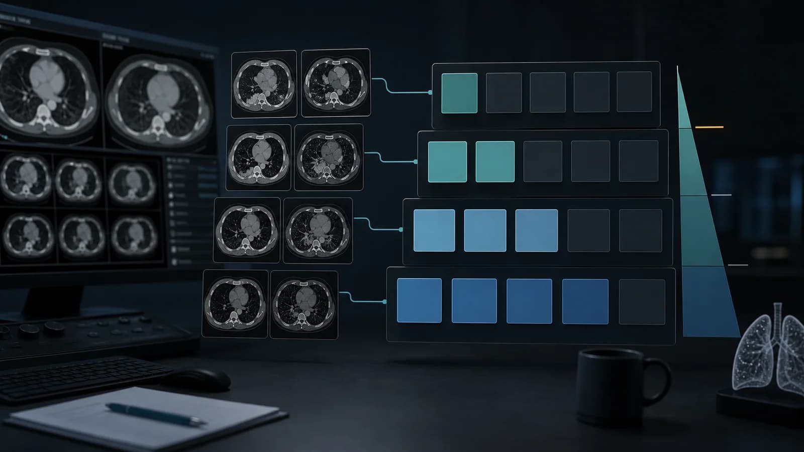
A subsolid pulmonary nodule is either pure ground glass or part solid. That first distinction changes the management pathway. Reliable classification and growth assessment require thin sections, appropriate window settings, and comparison with prior examinations.
Fleischner recommendations address incidentally detected nodules. Screening nodules, known cancer, immunosuppression, and some younger patients fall outside that framework. The report should state the clinical setting before attaching a follow-up recommendation.
Assessment framework
- Classify the nodule as pure ground glass or part solid on thin-section lung images.
- Use multiplanar images to avoid mistaking a vessel or bronchus for a solid component.
- Measure the total nodule and any solid component separately.
- Consider transient infection when deciding whether short-interval imaging is needed.
- Distinguish solitary from multiple subsolid nodules before recommending management.
What the report should contain
- State the lobe, segment, and relationship to the pleura.
- Describe margins, air bronchograms, and internal bubble-like lucencies when present.
- Report total diameter and solid-component diameter in the same section.
- Name the date and measurement method of the prior examination.
- Tie follow-up advice to patient context and the guideline being used.
Common errors
- Calling partial-volume opacity on thick sections a true solid component.
- Treating a one or two millimeter difference as automatic growth.
- Applying an incidental-nodule table directly to a screening examination.
Example report wording
Example impression: A 9 mm part-solid nodule in the right upper lobe has a 3 mm solid component. No prior examination is available. If the clinical setting fits incidental-nodule management, guideline-based imaging should confirm persistence. The final interval depends on patient risk and local practice.
The guide to adenocarcinoma arising in subsolid nodules and cystic airspaces covers suspicious morphologic change in more detail.
References
- Fleischner Society. Guidelines for Management of Incidental Pulmonary Nodules Detected on CT. Source (opens in a new tab)
- Fleischner Society. Recommendations for Measuring Pulmonary Nodules at CT. Source (opens in a new tab)
Clinical note: This material is for radiology education. Do not generate an automatic diagnosis or management decision without checking guideline scope, patient factors, and local policy.
Clinical use note
This content is educational and is not patient-specific medical advice. Every phrase, template, classification result, and recommendation must be verified and adapted by a physician using the complete examination, clinical context, current guidelines, and institutional protocol.
Move the nodule findings into a report draft
Open a RadPhrases report draft, adapt the morphology and measurements to the case, then verify every line against the images.
Related calculators
Browse the other checklists, phrase sets, and systematic approaches in this topic.