
Lung-RADS v2022: Nodule Categories, Growth, and Reporting Guide
Learn how Lung-RADS v2022 handles solid, part-solid, and ground-glass nodules, growth, categories 4A–4X, and practical reporting.
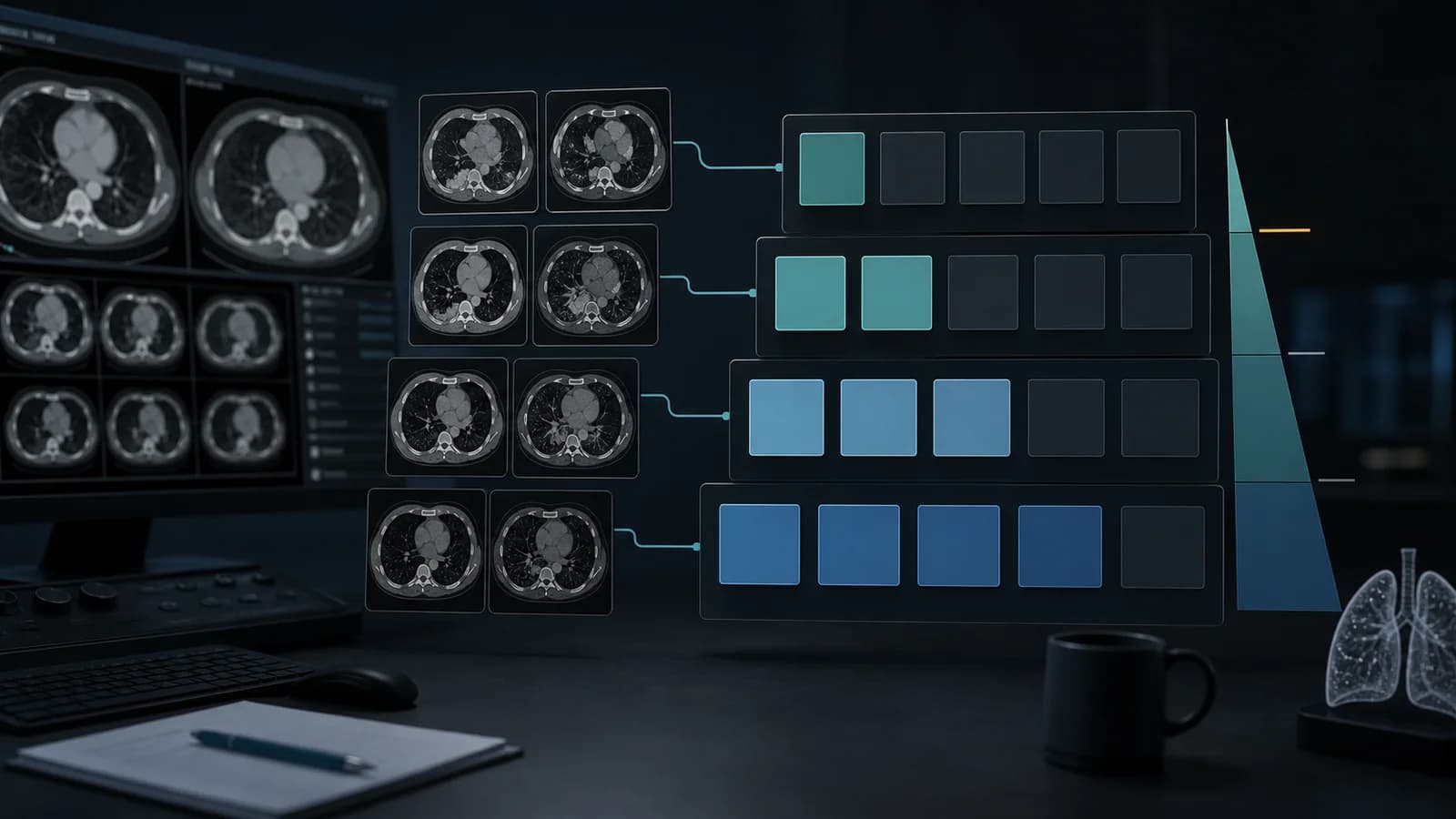
How to use Lung-RADS v2022
Lung-RADS v2022 assigns a screening examination category from its most suspicious eligible finding and links that category to a management pathway. It is intended for low-dose CT performed within a lung cancer screening program; do not automatically apply it to diagnostic chest CT in symptomatic patients or to incidentally detected nodules.
This article does not replace the official ACR Lung-RADS v2022 table. Every screening program should operate according to current national eligibility criteria and multidisciplinary protocols.
General meaning of the categories
| Category | General meaning | Typical next step |
|---|---|---|
| 0 | Incomplete or temporary assessment | Comparison with prior CT or short-interval reassessment |
| 1 | Negative | Screening LDCT in 12 months |
| 2 | Benign appearance or indolent behavior | Screening LDCT in 12 months |
| 3 | Probably benign | LDCT in 6 months |
| 4A | Suspicious | Usually LDCT in 3 months; PET/CT in selected cases |
| 4B | Very suspicious | Diagnostic CT, PET/CT, tissue sampling, or clinical evaluation |
| 4X | Category 3 or 4 nodule with additional suspicious features | Further evaluation according to the category and additional finding |
| S | Significant finding unrelated to lung cancer | Management appropriate to the finding |
The examination category is determined by the most suspicious nodule. Even when an examination is classified as negative, other nodules that do not alter the category may still be present.
1. Confirm that the examination is a screening study
Lung-RADS should be used only when the examination is part of a lung cancer screening program. A history of cancer, new hemoptysis, or unexplained weight loss may shift the examination into a diagnostic context. In such a setting, mechanically applying a screening table may fail to answer the clinical question.
2. Determine the nodule type
Place the nodule into one of the following groups:
- Solid nodule
- Part-solid nodule
- Non-solid/ground-glass nodule
- Juxtapleural nodule
- Airway nodule
- Atypical pulmonary cyst
Incorrectly selecting the nodule type changes the category thresholds entirely. In a part-solid nodule, measure and report the total diameter and the diameter of the solid component separately.
3. Standardize size measurement
In v2022, mean diameter may be reported to the nearest tenth of a millimeter as the average of the long- and short-axis measurements. When available, volume may also be reported and rounded to the nearest whole cubic millimeter.
For comparison, use the same window, similar section thickness, and a consistent measurement method. Thin-section reconstructions are particularly important when assessing the solid component of a part-solid nodule.
4. Define growth
Lung-RADS v2022 defines growth as an increase in mean diameter of more than 1.5 mm within a 12-month interval. However, solid or part-solid nodules that grow more slowly but continuously over serial examinations may still be suspicious. They should not be considered benign merely because they do not meet the single-year threshold.
Thresholds for a new nodule are not the same as those used for a nodule at baseline. A newly developed smaller nodule may enter a higher category.
5. Basic approach to solid nodules
The main thresholds in the official v2022 table can be summarized as follows:
- A solid nodule smaller than 6 mm at baseline is generally category 2.
- A solid nodule measuring 6 to <8 mm at baseline is category 3.
- A solid nodule measuring 8 to <15 mm at baseline is category 4A.
- A solid nodule measuring 15 mm or more at baseline is category 4B.
Lower diameter thresholds apply to new or growing nodules. The report should therefore state not only the current size, but also whether the nodule is new or growing.
6. Assess the solid component separately in part-solid nodules
In a part-solid nodule, the solid component has a major effect on malignancy risk in addition to the total diameter. At baseline, a nodule with a total diameter of 6 mm or more and a solid component smaller than 6 mm often enters the category 3 branch. A solid component measuring 6 to <8 mm raises consideration of category 4A; 8 mm or more raises consideration of category 4B.
Writing only “12 mm part-solid nodule” is insufficient. Record both measurements—for example, “total diameter 12 mm, solid component 5 mm.”
7. Consider behavior in non-solid nodules
Ground-glass nodules may follow a more indolent course. Under v2022, many non-solid nodules smaller than 30 mm remain category 2, whereas new or baseline nodules measuring 30 mm or more may enter category 3. A slowly enlarging non-solid nodule is managed according to the system's specific notes as long as no solid component develops.
The development of a new solid component during follow-up may be more important than an increase in total diameter.
8. Recognize juxtapleural and airway nodules
Small, smooth, oval, lentiform, or triangular nodules adjacent to the pleura and showing the typical appearance of a benign intrapulmonary lymph node may be category 2.
Segmental or more proximal airway nodules may be assigned category 4A at baseline. If a proximal airway nodule persists at 3-month follow-up, it may be upgraded to 4B, prompting consideration of additional evaluation such as bronchoscopy.
9. Do not overlook atypical pulmonary cysts
A major v2022 update is a more detailed approach to atypical pulmonary cysts. Thin-walled, regular cysts are generally not managed within Lung-RADS. Thick walls, asymmetric wall thickening, a mural nodule, multiloculation, or serial growth increase suspicion.
In a cavitary nodule, use the atypical-cyst approach when wall thickness is the dominant feature; use the solid-nodule approach when a nodular component is dominant.
10. Use category 4X correctly
Category 4X is used when a category 3 or 4 nodule has additional imaging features that increase suspicion for malignancy. Examples include:
- Spiculation
- Lymphadenopathy
- Findings of metastatic disease
- Rapid suspicious change
- Development of a new solid component in a growing non-solid nodule
4X is not an “X modifier”; it is a separate Lung-RADS category. It should not be confused with the S modifier.
What is the S modifier?
S may be added to categories 0–4 for a clinically significant or potentially significant finding not directly related to lung cancer. It may not be necessary for a stable finding that is already known and under evaluation. Coronary calcification, an aortic aneurysm, or another important finding should be reported according to the local protocol.
Example case 1: Baseline solid nodule
Findings: A solid nodule with a mean diameter of 7.2 mm in the right upper lobe on the initial screening LDCT.
- Baseline examination
- Solid nodule
- 6 to <8 mm
- Lung-RADS 3
Report example:
There is a solid pulmonary nodule in the right upper lobe with a mean diameter of 7.2 mm. The finding is consistent with Lung-RADS v2022 category 3. Follow-up with low-dose chest CT in 6 months is recommended.
Example case 2: Part-solid nodule
Findings: A baseline part-solid nodule in the left upper lobe with a total diameter of 14 mm and a 7 mm solid component.
- Baseline examination
- Solid component 6 to <8 mm
- Lung-RADS 4A
Report example:
In the left upper lobe, there is a part-solid nodule with a total mean diameter of 14 mm and a solid-component mean diameter of 7 mm. Lung-RADS v2022 category 4A. Low-dose chest CT in 3 months is recommended; PET/CT may be considered according to the size of the solid component and clinical risk.
Common mistakes made by residents
1. Using Lung-RADS on a non-screening CT
A different guideline may be appropriate for an incidental nodule or a symptomatic patient.
2. Applying baseline thresholds to a new nodule
New nodules may receive a higher category at a smaller diameter.
3. Reporting only the total diameter of a part-solid nodule
The solid component must be measured separately.
4. Using a single axis instead of mean diameter
Measure consistently using the average of the long and short axes.
5. Assigning the category according to the largest rather than the most suspicious nodule
Nodule type, growth, and additional suspicious features may be more important than size alone.
6. Automatically classifying an infectious appearance as 4B
An indeterminate inflammatory or infectious appearance may be assessed as Lung-RADS 0 with LDCT in 1–3 months in selected cases. A more malignant-appearing focus should receive the appropriate category.
Workstation checklist
- Is this truly a screening examination?
- Is prior CT available, and has it been compared?
- Is the nodule type correct?
- Was mean diameter or volume measured consistently?
- Was the solid component measured separately in a part-solid nodule?
- Is the nodule new, growing, or stable?
- Did I assign the category according to the most suspicious nodule?
- Did I distinguish 4X from the S modifier?
- Does the recommendation carry the same time interval as the category?
Frequently asked questions
Does Lung-RADS 4A mean cancer?
No. It is a suspicious-finding category and usually requires short-interval follow-up. PET/CT or additional evaluation may be added according to the clinical context.
Are the Fleischner criteria and Lung-RADS the same?
No. Lung-RADS is used for nodules detected within a screening program; Fleischner recommendations are generally used for incidentally detected nodules.
If a nodule grows by 1.4 mm in 12 months, does that mean there is no growth?
Although it does not meet the official growth threshold, continuous enlargement across multiple examinations may still be suspicious. Serial behavior should be assessed clinically, especially for solid or part-solid nodules.
References
- American College of Radiology. Lung CT Screening Reporting & Data System (Lung-RADS). Source (opens in a new tab)
- American College of Radiology. Lung-RADS v2022 Assessment Categories. Source (opens in a new tab)
- American College of Radiology. Summary of Changes from Lung-RADS v1.1 to v2022. ACR Lung-RADS resources.
Clinical use note
This content is educational and is not patient-specific medical advice. Every phrase, template, classification result, and recommendation must be verified and adapted by a physician using the complete examination, clinical context, current guidelines, and institutional protocol.
Build the category and report statement together
Use the relevant RadPhrases calculator, then verify the result against the official guideline and the complete examination.
Related calculators
Apply the classification or measurement steps from this guide in an interactive tool.