
Normal Noncontrast Head CT Report: Systematic Review and Sample Template
A systematic review sequence for noncontrast head CT, with a normal report example, short template, and commonly missed emergency findings.
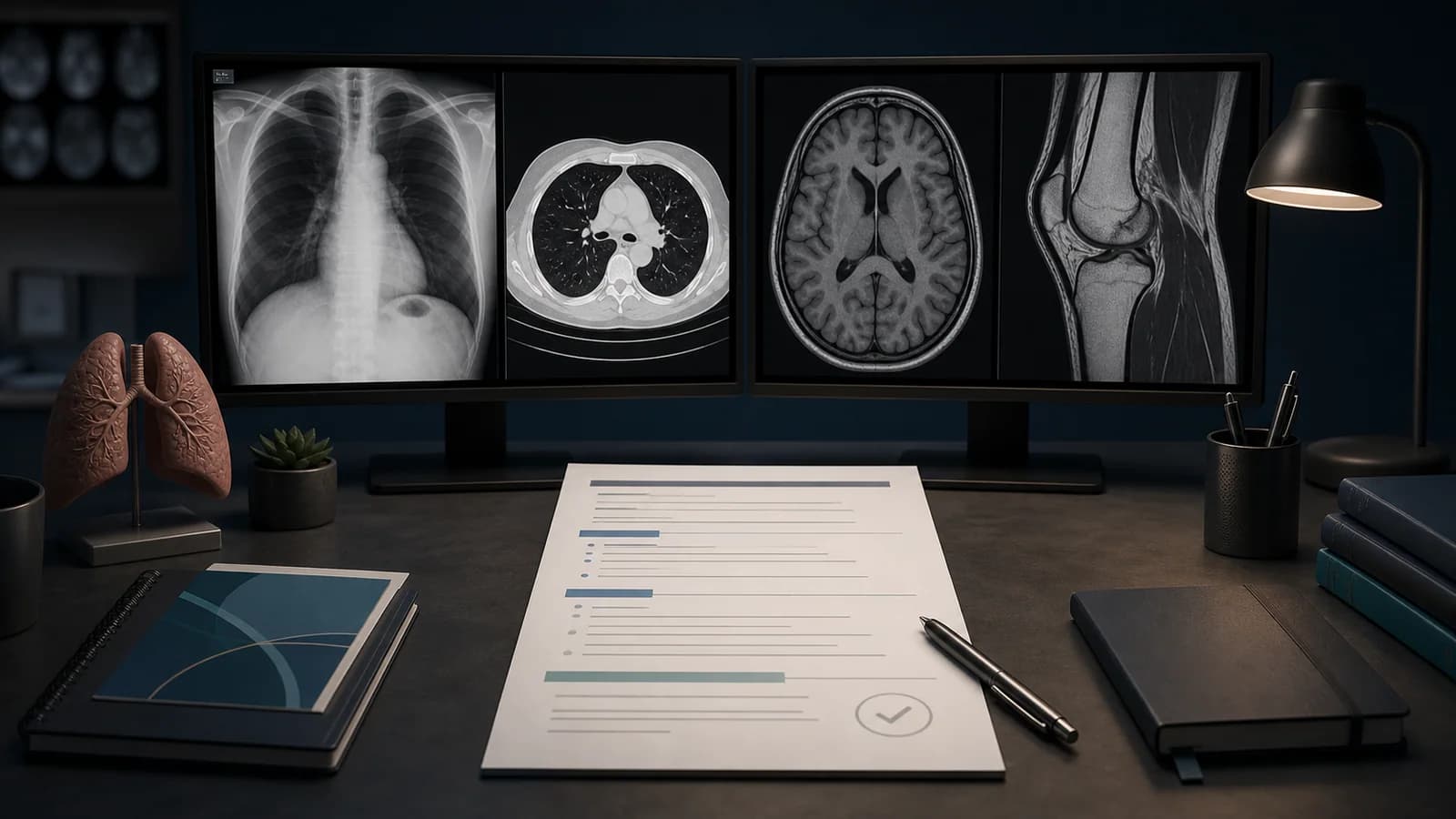
How to Write a Normal Noncontrast Head CT Report
Noncontrast head CT is one of the most common examinations in emergency radiology. Although the final report may be short, the study requires a systematic review for intracranial hemorrhage, early ischemic change, mass effect, hydrocephalus, and osseous abnormality.
A “normal head CT” does not always answer the entire clinical question. Early ischemia may be occult, while small posterior fossa lesions or subtle masses may be poorly assessed on noncontrast CT. The report should therefore reflect the indication and the limitations of the technique.
Clinical safety note: A normal template does not replace clinical assessment or MRI when clinically indicated. Every negative statement in the template must be independently verified on the images.
Pre-interpretation checks
Confirm the following:
- Is the study noncontrast or contrast-enhanced?
- What is the indication: trauma, headache, focal neurologic deficit, seizure, or altered mental status?
- Is prior neuroimaging available?
- Is there motion or metal artifact?
- Does the study adequately cover the skull base through the vertex?
- Were both brain and bone windows reviewed?
- Is there relevant history of surgery, shunt placement, or implants?
Systematic review sequence
1. Extra-axial spaces and hemorrhage
Review the convexities, fissures, cisterns, tentorium, and falx for subdural, epidural, and subarachnoid blood. Small hyperdense hemorrhages may be overlooked without window adjustment.
2. Brain parenchyma
Assess gray-white differentiation, symmetry, focal hypoattenuation or hyperattenuation, and intraparenchymal hemorrhage. In suspected acute ischemia, inspect the insular cortex, lentiform nuclei, and cortical sulci for early changes.
3. Mass effect and midline structures
Check for sulcal effacement, ventricular compression, herniation, and midline shift. “No mass effect” should reflect the effects of edema, hemorrhage, or collections, rather than only a visible mass.
4. Ventricular system and cisterns
Review the lateral, third, and fourth ventricles and the basal cisterns. Ventricular size should be interpreted in the context of age and prior imaging. Symmetric enlargement is not automatically physiologic.
5. Posterior fossa
The brainstem and cerebellum may be limited by beam-hardening artifact. Review for asymmetry, hemorrhage, mass effect, and fourth-ventricular distortion, and state the limitation when relevant.
6. Skull and extracranial structures
On bone windows, inspect the calvarium and skull base, particularly in trauma. Review the visible paranasal sinuses, mastoid air cells, orbits, and scalp soft tissues as appropriate to the indication.
Detailed normal noncontrast head CT example
Technique: Noncontrast head CT was performed with axial images and multiplanar reformations.

Comparison: None available.

Findings: No acute intracranial hemorrhage is identified. Gray-white matter differentiation is preserved. No focal parenchymal attenuation abnormality is identified. No mass effect or midline shift. The ventricles and cortical sulci are appropriate for age. The basal cisterns are patent. No acute calvarial fracture is identified. The visualized paranasal sinuses and mastoid air cells show no significant abnormality.

Impression: No acute intracranial abnormality.
The phrases “gray-white differentiation is preserved” and “no acute intracranial abnormality” do not completely exclude early ischemia.
Short report example
No acute intracranial hemorrhage, focal parenchymal attenuation abnormality, mass effect, or midline shift. Ventricular size is appropriate for age. No acute calvarial fracture.

Impression: No acute intracranial abnormality.
A short report does not mean that the sinuses, mastoids, or extracranial structures were ignored.
Adapting the template for trauma
No acute intracranial hemorrhage or mass effect. No acute fracture of the visualized calvarium.Do not use partial visualization of the upper cervical spine on head CT to declare the entire cervical spine normal.
Caution in acute neurologic deficit
One major purpose of noncontrast CT is to assess for hemorrhage and visible early ischemic change. A normal early study does not definitively exclude acute ischemia. Depending on timing and clinical suspicion, CTA, perfusion imaging, or MRI may be required under local protocols.
Common trainee mistakes
Skipping bone windows
The calvarium, skull base, and sinus walls should be reviewed even in the absence of known trauma.
Confusing age-related volume loss with hydrocephalus
Joint enlargement of the ventricles and sulci differs from disproportionate ventricular enlargement, sulcal effacement, or transependymal edema.
Treating the posterior fossa as automatically normal
CT sensitivity may be limited in this region. A technical limitation should be acknowledged when clinically relevant.
Converting sinus opacification directly into a clinical diagnosis
Mucosal thickening is an imaging observation, not a stand-alone diagnosis of clinical sinusitis.
Leaving “no acute abnormality” unchanged despite chronic findings
Chronic infarction, white-matter change, volume loss, or postoperative findings mean the study is not completely normal. The impression must match the actual findings.
Frequently asked questions
Does “no acute intracranial abnormality” exclude acute ischemia?
No. Early ischemia may be occult on noncontrast CT, especially soon after symptom onset.
Should every normal head CT report include the sinuses?
The visualized sinuses should be reviewed, but the level of detail should depend on the indication and clinical relevance. Nonvisualized regions should not be described as normal.
Does “age-appropriate volume loss” belong in a normal report?
If visible atrophy is present, the study is not strictly normal. Use careful and consistent terminology.
References
- American College of Radiology. ACR–ASNR–SPR Practice Parameter for the Performance of Computed Tomography (CT) of the Head.
- American College of Radiology. ACR Practice Parameter for Communication of Diagnostic Imaging Findings.
- RSNA. RadReport Template Library.
Legal and clinical note: This article is educational and does not replace diagnostic judgment, local protocols, or specialist review. Reports must be adapted to the case, image quality, and clinical question.
Clinical use note
This content is educational and is not patient-specific medical advice. Every phrase, template, classification result, and recommendation must be verified and adapted by a physician using the complete examination, clinical context, current guidelines, and institutional protocol.
Start from an editable report structure
Open a normal report framework in RadPhrases, then verify and adapt every line to the images and clinical question.
Related calculators
Browse the other checklists, phrase sets, and systematic approaches in this topic.