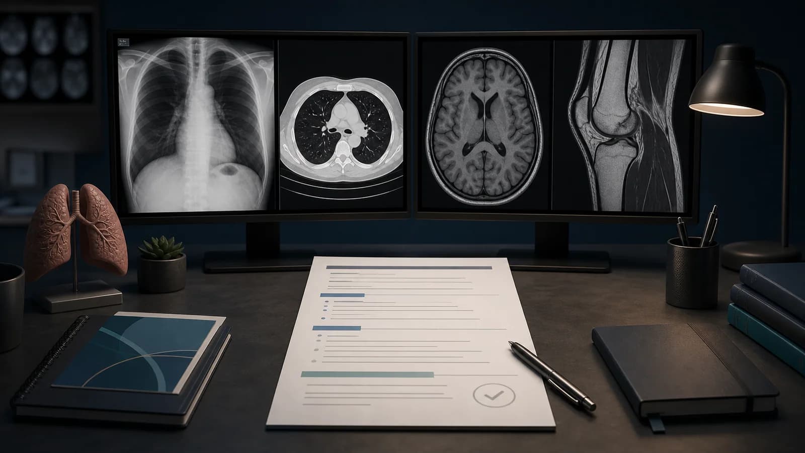
How to Write a Normal Chest Radiograph Report
A normal chest radiograph report may be brief, but brevity should not mean a superficial review. A reliable “normal” report requires confirmation of technical adequacy, followed by a fixed review of the cardiomediastinal structures, lungs, pleural spaces, and the visible bones and soft tissues.
The approach below is designed for radiology trainees, especially early in residency. The sample text below is educational and should be adapted to the patient’s age, projection, clinical indication, prior studies, and local terminology.
Clinical safety note: A normal template does not replace independent image interpretation. Only report a structure as negative when it was actually reviewed and adequately visualized.
Technical checks before calling the study normal
The first line of the report should make clear which images were reviewed. Upright PA and lateral views should not be interpreted in the same way as a portable AP study.
Check the following:
- Projection: PA, AP, lateral, or single view
- Position: upright, seated, or supine
- Rotation
- Degree of inspiration
- Penetration
- Inclusion of the apices and costophrenic angles
- Motion or external-object artifacts
- Availability of prior imaging
Remember that AP projection may magnify the cardiac silhouette, while small pneumothoraces or pleural effusions may have an atypical appearance on supine images. When technique is limited, state the limitation rather than using an unqualified normal template.
Systematic review sequence
1. Airway
Assess tracheal position and the patency of the main bronchi. Mild deviation should be interpreted in the context of rotation, anatomy, and the remainder of the examination.
Evaluate heart size in relation to the projection. Review mediastinal width, the aortic contour, and the hila. Measurement-based statements such as “normal cardiothoracic ratio” should be used cautiously outside an appropriate PA study.
3. Lung parenchyma
Compare both lungs from apex to base and from central to peripheral regions. Look for focal air-space opacity, interstitial opacity, nodules, volume loss, or asymmetry.
4. Pleura
Review the costophrenic angles, lateral pleural margins, and apices. Pleural effusion and pneumothorax should be checked separately from the lung parenchyma.
5. Diaphragm and upper abdomen
Assess both hemidiaphragms, the subdiaphragmatic regions, and the visible upper abdomen. Do not overlook incidental findings relevant to the clinical question.
6. Bones and soft tissues
Review the ribs, clavicles, shoulder girdles, thoracic spine, and chest-wall soft tissues to the extent permitted by the study. Although a chest radiograph is not a dedicated skeletal examination, obvious acute abnormalities should be reported.
7. Tubes, lines, and devices
On portable or postoperative studies, device assessment is a separate step. A study containing a central venous catheter, endotracheal tube, enteric tube, drain, or cardiac device should not be considered complete until device position has been evaluated.
Detailed normal PA and lateral chest radiograph example
Examination: PA and lateral chest radiographs.

Comparison: None available.

Findings: The cardiomediastinal silhouette is within normal limits in size and contour. No focal air-space opacity or significant interstitial abnormality is identified. No pleural effusion or pneumothorax is present. No acute osseous abnormality is identified on the included images.

Impression: No acute cardiopulmonary abnormality.
The phrase “no acute osseous abnormality” should only be used when the visible bones were adequately reviewed for that purpose.
Short normal report example
The cardiomediastinal silhouette is within normal limits. No focal air-space opacity is identified. No pleural effusion or pneumothorax.

Impression: No acute cardiopulmonary abnormality.
Some institutions use “no active infiltrate,” but the more observational phrase “no focal air-space opacity” may be clearer.
Example approach for a single-view AP study
A single AP chest radiograph was reviewed. The cardiomediastinal silhouette is suboptimally assessed because of AP technique. No focal air-space opacity is identified. No pleural effusion or pneumothorax.

Impression: No acute pulmonary parenchymal abnormality.
This wording should still be adapted for low lung volumes, rotation, supine positioning, or other technical limitations.
Common trainee mistakes
Calling cardiomegaly without considering projection
AP magnification may make the cardiac silhouette appear enlarged. Interpret heart size together with projection and inspiration.
Writing “lungs are normal” while skipping the pleura
The lungs may look clear while a small pneumothorax or pleural effusion is present. Pleural review should remain a distinct step.
Failing to mention technical limitations
Low lung volumes may accentuate basal linear opacities, while rotation can create hilar or mediastinal asymmetry. Check technique before converting an appearance into a diagnosis.
Ignoring bones and soft tissues
Rib fractures, shoulder abnormalities, subcutaneous emphysema, or breast-shadow asymmetry may be visible on a chest radiograph.
Writing an impression that does not answer the clinical question
For a patient with fever and cough, the impression should clearly state whether a focal opacity concerning for pneumonia is present.
Frequently asked questions
Is “no infiltrate” enough for a normal chest radiograph?
Usually not. Pleural effusion, pneumothorax, cardiomediastinal appearance, and other visible structures should also be assessed.
Should a normal report mention comparison?
If a prior examination is available and clinically relevant, comparison is useful. If no prior study exists, the report may state “none available,” depending on local practice.
Should every normal study include a negative bone statement?
Not necessarily. The visible bones and soft tissues should still be reviewed. Any negative statement should remain within the actual scope of the examination.
References
- American College of Radiology. ACR–SPR–STR Practice Parameter for the Performance of Chest Radiography.
Source (opens in a new tab)
- American College of Radiology. ACR Practice Parameter for Communication of Diagnostic Imaging Findings.
Source (opens in a new tab)
- RSNA. RadReport Template Library.
Source (opens in a new tab)
Legal and clinical note: This article is educational and does not replace medical advice, local protocols, or specialist judgment. Sample reports must be adapted to the actual patient, images, and clinical context.