
Normal Noncontrast Chest CT Report: Systematic Review and Sample Template
A systematic approach to the lungs, airways, pleura, mediastinum, bones, and upper abdomen on noncontrast chest CT, with a normal report example.
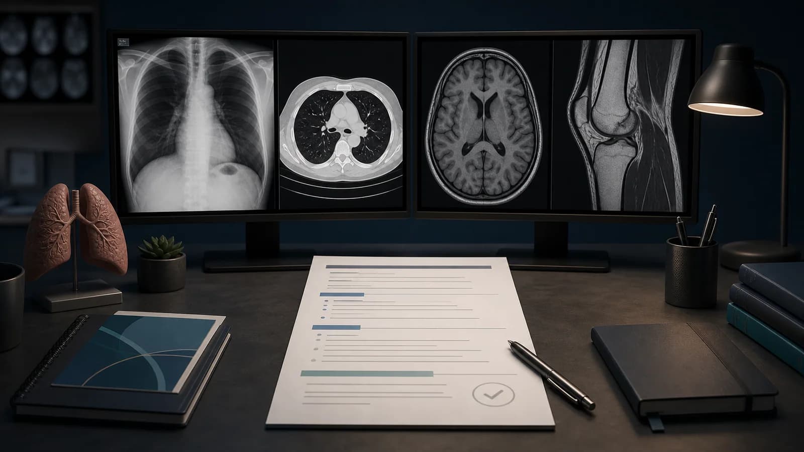
How to Write a Normal Noncontrast Chest CT Report
A normal chest CT report should include more than the statement “the lungs are clear.” Chest CT covers the lung parenchyma, airways, pleura, mediastinum, assessable aspects of the heart and great vessels, lymph nodes, chest wall, bones, and visible upper abdomen.
Report content varies with protocol. Standard-dose CT, low-dose screening CT, and high-resolution CT should not share an identical template. Noncontrast chest CT is not an appropriate examination for excluding pulmonary embolism, and the report should not make negative vascular claims beyond the protocol.
Clinical safety note: A normal template does not expand the diagnostic scope of the protocol. Do not report a condition as absent when the examination was not designed to assess it.
Pre-interpretation checks
- Is the study noncontrast or contrast-enhanced?
- Is it standard chest CT, low-dose screening, HRCT, or another protocol?
- Is inspiration adequate?
- Is there respiratory or motion artifact?
- Were lung and mediastinal windows both reviewed?
- Were coronal and sagittal reformations reviewed?
- Is prior imaging available?
- Is the clinical question infection, nodule, interstitial disease, trauma, or malignancy follow-up?
Systematic review sequence
1. Lung parenchyma
Review both lungs by lobe and segment from apex to base. Look for focal consolidation, ground-glass opacity, nodule, mass, atelectasis, emphysema, and interstitial change.
The phrase “no pulmonary nodule” requires technically appropriate imaging and careful review of the entire parenchyma.
2. Central airways
Assess the trachea and main bronchi for patency, wall abnormality, or endobronchial lesion. Expiratory collapse and small-airway disease may be limited on a routine inspiratory study.
3. Pleura
Review for pleural effusion, pneumothorax, pleural thickening, and plaque. Very small effusions may collect in the posterior recesses.
4. Mediastinum and hila
Assess mediastinal and hilar lymph nodes, masses, and fat planes. On noncontrast CT, distinguishing hilar vessels from lymph nodes may be limited.
5. Heart and great vessels
Heart size, pericardial fluid, and thoracic aortic caliber may be assessed. Luminal disease, dissection, embolism, or vascular-wall pathology requires the appropriate protocol.
6. Chest wall, axillae, and visible thyroid
Review the axillary nodes, chest-wall soft tissues, and visible thyroid for significant abnormalities. Chest CT does not replace dedicated breast or thyroid imaging.
7. Bones
Review the ribs, sternum, vertebrae, and shoulder girdles on bone windows. Do not overlook fracture, destructive lesions, or relevant degenerative change.
8. Upper abdomen
Inspect the visible portions of the liver, spleen, adrenal glands, and kidneys. Partial visualization should not be summarized as a normal abdominal examination.
Detailed normal noncontrast chest CT example
Technique: Noncontrast CT of the chest was performed with axial images and multiplanar reformations.

Comparison: None available.

Findings: The trachea and central bronchi are patent. No focal consolidation, significant ground-glass opacity, suspicious pulmonary nodule, or mass is identified. No pleural effusion or pneumothorax. No pathologically enlarged mediastinal or hilar lymph node is identified. Heart size is within normal limits. No pericardial effusion. The thoracic aorta is normal in caliber. No significant abnormality is identified in the visualized upper abdomen. No acute or destructive osseous abnormality is identified.

Impression: No significant intrathoracic abnormality on noncontrast chest CT.
The phrase “no suspicious pulmonary nodule” should only be used when image quality and reconstruction thickness are suitable.
Short normal report example
No focal consolidation, suspicious pulmonary nodule, or mass. The central airways are patent. No pleural or pericardial effusion. No pathologically enlarged mediastinal lymph node.

Impression: No significant intrathoracic abnormality.
A short report still requires review of the bones, chest wall, and visible upper abdomen.
Why HRCT needs a separate template
High-resolution CT for interstitial lung disease evaluates reticulation, traction bronchiectasis, honeycombing, air trapping, and disease distribution. Inspiratory, expiratory, or prone images must be incorporated when obtained.
A routine noncontrast chest CT template should therefore not be copied directly into an HRCT report.
How the template changes after contrast
Contrast-enhanced studies improve evaluation of vessels and mediastinal structures, but timing remains critical. A routine venous-phase chest CT is not equivalent to CT pulmonary angiography.
Common trainee mistakes
Reading only the lung windows
Lymph nodes, pericardial fluid, aortic caliber, and mediastinal masses require mediastinal-window review.
Excluding pulmonary embolism on noncontrast CT
Pulmonary arterial lumina cannot be reliably assessed without the appropriate contrast-enhanced angiographic protocol.
Ignoring the upper abdomen
Important adrenal or hepatic findings may appear on chest CT.
Treating every lymph node as pathologic
Size, morphology, distribution, and clinical context should be interpreted together.
Adding unnecessary certainty
When image quality or protocol is limited, use a conclusion that accurately defines the scope rather than “no abnormality whatsoever.”
Frequently asked questions
Does a normal noncontrast chest CT exclude pulmonary embolism?
No. Pulmonary embolism assessment requires a properly timed CT pulmonary angiography protocol.
Should every normal chest CT include a negative statement about nodules?
Only when image quality and technique allow a confident assessment. Avoid overly definitive wording in the presence of thick sections, motion, or other limitations.
Should upper-abdominal findings be included in a chest CT report?
Clinically significant findings in the visible portions should be reported, while recognizing that partial imaging does not replace a dedicated abdominal examination.
References
- American College of Radiology. ACR–SABI–SPR–STR Practice Parameter for the Performance of Thoracic Computed Tomography (CT).
- American College of Radiology. ACR Practice Parameter for Communication of Diagnostic Imaging Findings.
- RSNA. RadReport Template Library.
Legal and clinical note: This article is educational and does not replace patient-specific assessment, current guidelines, local protocols, or specialist judgment.
Clinical use note
This content is educational and is not patient-specific medical advice. Every phrase, template, classification result, and recommendation must be verified and adapted by a physician using the complete examination, clinical context, current guidelines, and institutional protocol.
Start from an editable report structure
Open a normal report framework in RadPhrases, then verify and adapt every line to the images and clinical question.
Related calculators
Browse the other checklists, phrase sets, and systematic approaches in this topic.